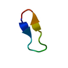
SEQUENCE THIS IS PART OF AN OPTIMIZED BETA-HAIRPIN SCAFFOLD, (SEE RUSSELL, BLANDL, SKELTON, & ...SEQUENCE THIS IS PART OF AN OPTIMIZED BETA-HAIRPIN SCAFFOLD, (SEE RUSSELL, BLANDL, SKELTON, & COCHRAN, "STABILITY OF CYCLIC BETA-HAIRPINS: ASYMMETRIC CONTRIBUTIONS FROM SIDE CHAINS OF A HYDROGEN-BONDED CROSS-STRAND RESIDUE PAIR", J. AMER. CHEM. SOC., IN PRESS.)
BLySReceptor3 / Tumor necrosis factor receptor superfamily member 13C / B cell-activating factor receptor / BAFF ...Tumor necrosis factor receptor superfamily member 13C / B cell-activating factor receptor / BAFF receptor / BAFF-R / BLys receptor 3
Mass: 1593.918 Da / Num. of mol.: 1 / Fragment: BR3 loop (residues 26-31) / Source method: obtained synthetically Details: The Peptide was synthesized chemically with acetylated N-terminus and amidated C-terminus References: UniProt: Q96RJ3
Has protein modification
Y
-
Experimental details
-
Experiment
Experiment
Method: SOLUTION NMR
NMR experiment
Conditions-ID
Experiment-ID
Solution-ID
Type
1
1
1
2D NOESY
1
2
1
2D TOCSY
1
3
1
DQF-COSY
1
4
2
2D NOESY
1
5
2
COSY-35
-
Sample preparation
Details
Solution-ID
Contents
Solvent system
1
2.9mMbhpBR3peptide
92% H2O, 8% D2O, 0.1mMDSS, pH4.5
2
2.9mMbhpBR3peptide
100% D2O, 0.1mMDSS, pH4.5
Sample conditions
Ionic strength: no added salt / pH: 4.5 / Pressure: ambient / Temperature: 293 K
Crystal grow
*PLUS
Method: other / Details: NMR
-
NMR measurement
Radiation
Protocol: SINGLE WAVELENGTH / Monochromatic (M) / Laue (L): M
Radiation wavelength
Relative weight: 1
NMR spectrometer
Type: Bruker DRX / Manufacturer: Bruker / Model: DRX / Field strength: 600 MHz
-
Processing
NMR software
Name
Version
Developer
Classification
XwinNMR
1.37.4.2
Bruker
collection
Felix
98
Accelrys
dataanalysis
DGII
98
Accelrys
structuresolution
Discover
98
Accelrys
refinement
Refinement
Method: hybrid distance geometry, simulated annealing, restrained molecular dynamics. Software ordinal: 1 Details: Complete 1H resonance assignments were obtained using standard 2D homonuclear NMR methods. Distance restraints were derived from analysis of a 2D NOESY spectrum (250ms mixing time); HN-Ha ...Details: Complete 1H resonance assignments were obtained using standard 2D homonuclear NMR methods. Distance restraints were derived from analysis of a 2D NOESY spectrum (250ms mixing time); HN-Ha coupling constants were obtained from analysis of a DQF-COSY spectrum acquired in water; and HaHb values were obtained from analysis of a COSY-35 spectrum acquired in D2O. Structures were calculated from a total of 119 NOE-derived (including 46 long-range) distance restraints and 16 dihedral angle restraints. Structures satisfy the experimental data very well with no distance or dihedral angle violations greater than 0.1 angstrom or 1 degree, respectively.
NMR representative
Selection criteria: closest to the average
NMR ensemble
Conformer selection criteria: structures with the least restraint violations Conformers calculated total number: 80 / Conformers submitted total number: 20
+
About Yorodumi
-
News
-
Feb 9, 2022. New format data for meta-information of EMDB entries
New format data for meta-information of EMDB entries
Version 3 of the EMDB header file is now the official format.
The previous official version 1.9 will be removed from the archive.
In the structure databanks used in Yorodumi, some data are registered as the other names, "COVID-19 virus" and "2019-nCoV". Here are the details of the virus and the list of structure data.
Jan 31, 2019. EMDB accession codes are about to change! (news from PDBe EMDB page)
EMDB accession codes are about to change! (news from PDBe EMDB page)
The allocation of 4 digits for EMDB accession codes will soon come to an end. Whilst these codes will remain in use, new EMDB accession codes will include an additional digit and will expand incrementally as the available range of codes is exhausted. The current 4-digit format prefixed with “EMD-” (i.e. EMD-XXXX) will advance to a 5-digit format (i.e. EMD-XXXXX), and so on. It is currently estimated that the 4-digit codes will be depleted around Spring 2019, at which point the 5-digit format will come into force.
The EM Navigator/Yorodumi systems omit the EMD- prefix.
Related info.:Q: What is EMD? / ID/Accession-code notation in Yorodumi/EM Navigator
Yorodumi is a browser for structure data from EMDB, PDB, SASBDB, etc.
This page is also the successor to EM Navigator detail page, and also detail information page/front-end page for Omokage search.
The word "yorodu" (or yorozu) is an old Japanese word meaning "ten thousand". "mi" (miru) is to see.
Related info.:EMDB / PDB / SASBDB / Comparison of 3 databanks / Yorodumi Search / Aug 31, 2016. New EM Navigator & Yorodumi / Yorodumi Papers / Jmol/JSmol / Function and homology information / Changes in new EM Navigator and Yorodumi
 Movie
Movie Controller

 Yorodumi
 Open data
 Basic information
 Components
 Keywords
 Function and homology information
 Authors
 Citation
 Structure visualization
 Downloads & links
 Other downloads
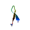


 PDBj
PDBj Assembly
 Sample preparation
 Processing