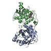
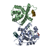
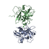
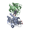
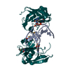
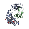
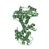
- PDB-1mkz: Crystal structure of MoaB protein at 1.6 A resolution. -
+
Open data
ID or keywords:
Loading...
-
Basic information
Entry
Database: PDB / ID: 1mkz
Title
Crystal structure of MoaB protein at 1.6 A resolution.
Components
Molybdenum cofactor biosynthesis protein B
Keywords
BIOSYNTHETIC PROTEIN / MAD / Weak anomalous signal / Molybdopterin synthesis / Structural Genomics / PSI / Protein Structure Initiative / Midwest Center for Structural Genomics / MCSG
Function / homology
Function and homology information
Mo-molybdopterin cofactor biosynthetic process / protein hexamerization / GTP binding / identical protein binding / cytosol Similarity search - Function
Highest resolution: 1.6 Å / Lowest resolution: 129.1 Å / Num. obs: 55534 / % possible obs: 99.4 % / Num. measured all: 311387 / Rmerge(I) obs: 0.057
Reflection shell
*PLUS
% possible obs: 99 % / Rmerge(I) obs: 0.45 / Mean I/σ(I) obs: 2.5
-
Processing
Software
Name
Version
Classification
REFMAC
5.1.24
refinement
HKL-2000
datareduction
CNS
refinement
WARP
modelbuilding
d*TREK
datascaling
d*TREK
datareduction
HKL-2000
datascaling
CNS
phasing
ARP/wARP
modelbuilding
Refinement
Resolution: 1.6→129.1 Å / Cor.coef. Fo:Fc: 0.962 / Cor.coef. Fo:Fc free: 0.952 / SU B: 1.703 / SU ML: 0.06 / Cross valid method: THROUGHOUT / ESU R: 0.095 / ESU R Free: 0.096 / Stereochemistry target values: MAXIMUM LIKELIHOOD / Details: HYDROGENS HAVE BEEN ADDED IN THE RIDING POSITIONS
Rfactor
Num. reflection
% reflection
Selection details
Rfree
0.21871
2248
5.1 %
RANDOM
Rwork
0.18284
-
-
-
obs
0.18461
42215
94.81 %
-
Solvent computation
Ion probe radii: 0.8 Å / Shrinkage radii: 0.8 Å / VDW probe radii: 1.4 Å / Solvent model: BABINET MODEL WITH MASK
Displacement parameters
Biso mean: 14.768 Å2
Baniso -1
Baniso -2
Baniso -3
1-
-0.04 Å2
-0.02 Å2
0 Å2
2-
-
-0.04 Å2
0 Å2
3-
-
-
0.05 Å2
Refinement step
Cycle: LAST / Resolution: 1.6→129.1 Å
Protein
Nucleic acid
Ligand
Solvent
Total
Num. atoms
2679
0
35
247
2961
Refine LS restraints
Refine-ID
Type
Dev ideal
Dev ideal target
Number
X-RAY DIFFRACTION
r_bond_refined_d
0.02
0.021
2762
X-RAY DIFFRACTION
r_bond_other_d
0.001
0.02
2551
X-RAY DIFFRACTION
r_angle_refined_deg
1.885
1.955
3754
X-RAY DIFFRACTION
r_angle_other_deg
0.856
3
5920
X-RAY DIFFRACTION
r_dihedral_angle_1_deg
6.285
5
339
X-RAY DIFFRACTION
r_dihedral_angle_2_deg
X-RAY DIFFRACTION
r_chiral_restr
0.107
0.2
443
X-RAY DIFFRACTION
r_gen_planes_refined
0.007
0.02
3019
X-RAY DIFFRACTION
r_gen_planes_other
0.003
0.02
537
X-RAY DIFFRACTION
r_nbd_refined
0.24
0.3
539
X-RAY DIFFRACTION
r_nbd_other
0.266
0.3
2945
X-RAY DIFFRACTION
r_nbtor_other
0.091
0.5
1627
X-RAY DIFFRACTION
r_xyhbond_nbd_refined
0.171
0.5
306
X-RAY DIFFRACTION
r_xyhbond_nbd_other
X-RAY DIFFRACTION
r_symmetry_vdw_refined
0.198
0.3
33
X-RAY DIFFRACTION
r_symmetry_vdw_other
0.329
0.3
109
X-RAY DIFFRACTION
r_symmetry_hbond_refined
0.278
0.5
22
X-RAY DIFFRACTION
r_symmetry_hbond_other
X-RAY DIFFRACTION
r_mcbond_it
0.886
1.5
1719
X-RAY DIFFRACTION
r_mcangle_it
1.487
2
2795
X-RAY DIFFRACTION
r_scbond_it
2.595
3
1043
X-RAY DIFFRACTION
r_scangle_it
3.641
4.5
959
X-RAY DIFFRACTION
r_rigid_bond_restr
X-RAY DIFFRACTION
r_sphericity_free
X-RAY DIFFRACTION
r_sphericity_bonded
LS refinement shell
Resolution: 1.6→1.642 Å / Total num. of bins used: 20 /
In the structure databanks used in Yorodumi, some data are registered as the other names, "COVID-19 virus" and "2019-nCoV". Here are the details of the virus and the list of structure data.
Jan 31, 2019. EMDB accession codes are about to change! (news from PDBe EMDB page)
EMDB accession codes are about to change! (news from PDBe EMDB page)
The allocation of 4 digits for EMDB accession codes will soon come to an end. Whilst these codes will remain in use, new EMDB accession codes will include an additional digit and will expand incrementally as the available range of codes is exhausted. The current 4-digit format prefixed with “EMD-” (i.e. EMD-XXXX) will advance to a 5-digit format (i.e. EMD-XXXXX), and so on. It is currently estimated that the 4-digit codes will be depleted around Spring 2019, at which point the 5-digit format will come into force.
The EM Navigator/Yorodumi systems omit the EMD- prefix.
Related info.:Q: What is EMD? / ID/Accession-code notation in Yorodumi/EM Navigator
Yorodumi is a browser for structure data from EMDB, PDB, SASBDB, etc.
This page is also the successor to EM Navigator detail page, and also detail information page/front-end page for Omokage search.
The word "yorodu" (or yorozu) is an old Japanese word meaning "ten thousand". "mi" (miru) is to see.
Related info.:EMDB / PDB / SASBDB / Comparison of 3 databanks / Yorodumi Search / Aug 31, 2016. New EM Navigator & Yorodumi / Yorodumi Papers / Jmol/JSmol / Function and homology information / Changes in new EM Navigator and Yorodumi
 Movie
Movie Controller


 Open data
 Basic information
 Components
 Keywords
 Function and homology information
 X-RAY DIFFRACTION /
  Authors
 Citation
 Structure visualization
 Downloads & links
 Other downloads
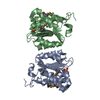


 PDBj
PDBj

 Assembly
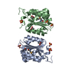

 Mass: 96.063 Da / Num. of mol.: 7 / Source method: obtained synthetically / Formula: SO4
 Mass: 60.052 Da / Num. of mol.: 3 / Source method: obtained synthetically / Formula: C2H4O2
 Mass: 18.015 Da / Num. of mol.: 235 / Source method: isolated from a natural source / Formula: H2O
 Sample preparation
 / Beamline: 19-BM
 Processing