 Movie
Movie Controller

[English] 日本語
 Yorodumi
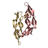
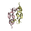
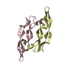
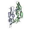
- PDB-1mkk: Disulfide deficient mutant of vascular endothelial growth factor ... -
+ Open data

- Basic information
| Entry | Database: PDB / ID: 1mkk | ||||||
|---|---|---|---|---|---|---|---|
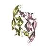
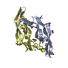
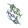
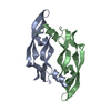
| Title | Disulfide deficient mutant of vascular endothelial growth factor A (C61A and C104A) | ||||||
 Components Components | Vascular Endothelial Growth Factor A | ||||||
 Keywords Keywords | HORMONE/GROWTH FACTOR / Cystine-knot growth factor / HORMONE-GROWTH FACTOR COMPLEX | ||||||
| Function / homology |  Function and homology information Function and homology informationSignaling by VEGF / basophil chemotaxis / positive regulation of endothelial cell chemotaxis by VEGF-activated vascular endothelial growth factor receptor signaling pathway / VEGF-A complex / cellular stress response to acid chemical / positive regulation of lymphangiogenesis / VEGF ligand-receptor interactions / vascular endothelial growth factor receptor 1 binding / negative regulation of adherens junction organization / primitive erythrocyte differentiation ...Signaling by VEGF / basophil chemotaxis / positive regulation of endothelial cell chemotaxis by VEGF-activated vascular endothelial growth factor receptor signaling pathway / VEGF-A complex / cellular stress response to acid chemical / positive regulation of lymphangiogenesis / VEGF ligand-receptor interactions / vascular endothelial growth factor receptor 1 binding / negative regulation of adherens junction organization / primitive erythrocyte differentiation / positive regulation of mast cell chemotaxis / negative regulation of establishment of endothelial barrier / vascular endothelial growth factor receptor binding / post-embryonic camera-type eye development / lymph vessel morphogenesis / negative regulation of blood-brain barrier permeability / positive regulation of cell proliferation by VEGF-activated platelet derived growth factor receptor signaling pathway / : / VEGF-activated neuropilin signaling pathway / bone trabecula formation / coronary vein morphogenesis / cardiac vascular smooth muscle cell development / lymphangiogenesis / motor neuron migration / VEGF binds to VEGFR leading to receptor dimerization / vascular endothelial growth factor receptor-2 signaling pathway / lung vasculature development / regulation of hematopoietic progenitor cell differentiation / eye photoreceptor cell development / positive regulation of axon extension involved in axon guidance / endothelial cell chemotaxis / positive regulation of protein localization to early endosome / positive regulation of protein autophosphorylation / positive regulation of trophoblast cell migration / vascular wound healing / positive regulation of epithelial tube formation / camera-type eye morphogenesis / neuropilin binding / positive regulation of blood vessel endothelial cell proliferation involved in sprouting angiogenesis / positive regulation of branching involved in ureteric bud morphogenesis / induction of positive chemotaxis / transmembrane receptor protein tyrosine kinase activator activity / coronary artery morphogenesis / negative regulation of cell-cell adhesion mediated by cadherin / tube formation / vascular endothelial growth factor receptor 2 binding / dopaminergic neuron differentiation / positive regulation of vascular permeability / commissural neuron axon guidance / positive regulation of vascular endothelial growth factor signaling pathway / positive regulation of peptidyl-tyrosine phosphorylation / cell migration involved in sprouting angiogenesis / positive regulation of blood vessel branching / surfactant homeostasis / platelet-derived growth factor receptor binding / epithelial cell maturation / sprouting angiogenesis / extracellular matrix binding / endothelial cell proliferation / positive regulation of positive chemotaxis / Regulation of gene expression by Hypoxia-inducible Factor / positive regulation of leukocyte migration / retinal ganglion cell axon guidance / positive regulation of endothelial cell chemotaxis / artery morphogenesis / cardiac muscle cell development / positive regulation of cell migration involved in sprouting angiogenesis / positive regulation of DNA biosynthetic process / negative regulation of epithelial to mesenchymal transition / vascular endothelial growth factor signaling pathway / branching involved in blood vessel morphogenesis / positive regulation of neuroblast proliferation / positive chemotaxis / negative regulation of fat cell differentiation / positive regulation of sprouting angiogenesis / chemoattractant activity / mesoderm development / outflow tract morphogenesis / fibronectin binding / positive regulation of cell division / macrophage differentiation / neuroblast proliferation / mammary gland alveolus development / cellular response to vascular endothelial growth factor stimulus / positive regulation of receptor internalization / positive regulation of focal adhesion assembly / monocyte differentiation / positive regulation of blood vessel endothelial cell migration / vascular endothelial growth factor receptor signaling pathway / positive regulation of osteoblast differentiation / vasculogenesis / ovarian follicle development / heart morphogenesis / lactation / TFAP2 (AP-2) family regulates transcription of growth factors and their receptors / cell maturation / epithelial cell differentiation / positive regulation of endothelial cell proliferation / lung development / positive regulation of endothelial cell migration Similarity search - Function | ||||||
| Biological species |  Homo sapiens (human) Homo sapiens (human) | ||||||
| Method |  X-RAY DIFFRACTION / SYNCHROTRON / MOLECULAR REPLACEMENT / Resolution: 1.32 Å X-RAY DIFFRACTION / SYNCHROTRON / MOLECULAR REPLACEMENT / Resolution: 1.32 Å | ||||||
 Authors Authors | Muller, Y.A. / Heiring, C. / Misselwitz, R. / Welfle, K. / Welfle, H. | ||||||
 Citation Citation | Journal: J.Biol.Chem. / Year: 2002 Title: The cystine knot promotes folding and not thermodynamic stability in vascular endothelial growth factor Authors: Muller, Y.A. / Heiring, C. / Misselwitz, R. / Welfle, K. / Welfle, H. #1: Journal: Protein Eng. / Year: 2001Title: Folding Screening Assayed By Proteolysis: Application to Various Cystine Deletion Mutants of VasculaR Endothelial Growth Factor Authors: Heiring, C. / Muller, Y.A. | ||||||
| History |
|
- Structure visualization
| Structure viewer | Molecule: MolmilJmol/JSmol |
|---|
- Downloads & links
-Download
| PDBx/mmCIF format | 1mkk.cif.gz | 96.3 KB | Display | PDBx/mmCIF format |
|---|---|---|---|---|
| PDB format | pdb1mkk.ent.gz | 74.5 KB | Display | PDB format |
| PDBx/mmJSON format | 1mkk.json.gz | Tree view | PDBx/mmJSON format | |
| Others |  Other downloads Other downloads |
-Validation report
| Arichive directory | https://data.pdbj.org/pub/pdb/validation_reports/mk/1mkkftp://data.pdbj.org/pub/pdb/validation_reports/mk/1mkk | HTTPS FTP |
|---|
-Related structure data
| Related structure data |  1mjvC  1mkgC  2vpfS S: Starting model for refinement C: citing same article ( |
|---|---|
| Similar structure data |

-Links
 PDBj
PDBj


- Assembly
| Deposited unit | 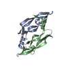
| ||||||||
|---|---|---|---|---|---|---|---|---|---|
| 1 |
| ||||||||
| Unit cell |
|
-Components
| #1: Protein | Mass: 11195.966 Da / Num. of mol.: 2 / Fragment: Residues 40-134, sequence database / Mutation: C61A,C104A Source method: isolated from a genetically manipulated source Source: (gene. exp.) Homo sapiens (human) / Plasmid: pET-3d / Production host:  #2: Water | ChemComp-HOH / |  Mass: 18.015 Da / Num. of mol.: 205 / Source method: isolated from a natural source / Formula: H2O Mass: 18.015 Da / Num. of mol.: 205 / Source method: isolated from a natural source / Formula: H2OHas protein modification | Y | |
|---|
-Experimental details
-Experiment
| Experiment | Method: X-RAY DIFFRACTION / Number of used crystals: 1 |
|---|
- Sample preparation
| Crystal | Density Matthews: 2.6 Å3/Da / Density % sol: 52.67 % | ||||||||||||||||||||||||||||||||||||||||||||||||||||||||||||
|---|---|---|---|---|---|---|---|---|---|---|---|---|---|---|---|---|---|---|---|---|---|---|---|---|---|---|---|---|---|---|---|---|---|---|---|---|---|---|---|---|---|---|---|---|---|---|---|---|---|---|---|---|---|---|---|---|---|---|---|---|---|
| Crystal grow | Temperature: 293 K / Method: vapor diffusion, hanging drop / pH: 5.6 Details: isopropanol, PEG4000, pH 5.6, VAPOR DIFFUSION, HANGING DROP, temperature 293K | ||||||||||||||||||||||||||||||||||||||||||||||||||||||||||||
| Crystal grow | *PLUS | ||||||||||||||||||||||||||||||||||||||||||||||||||||||||||||
| Components of the solutions | *PLUS
|
-Data collection
| Diffraction | Mean temperature: 100 K |
|---|---|
| Diffraction source | Source: SYNCHROTRON / Site: EMBL/DESY, HAMBURG  / Beamline: BW7B / Wavelength: 0.8423 Å / Beamline: BW7B / Wavelength: 0.8423 Å |
| Detector | Type: MARRESEARCH / Detector: IMAGE PLATE / Date: Aug 5, 2002 |
| Radiation | Protocol: SINGLE WAVELENGTH / Monochromatic (M) / Laue (L): M / Scattering type: x-ray |
| Radiation wavelength | Wavelength: 0.8423 Å / Relative weight: 1 |
| Reflection | Resolution: 1.32→28 Å / Num. all: 53321 / Num. obs: 53321 / % possible obs: 99.2 % / Observed criterion σ(F): 0 / Observed criterion σ(I): -3 / Redundancy: 3.8 % / Biso Wilson estimate: 22.06 Å2 / Rmerge(I) obs: 0.074 / Net I/σ(I): 12.08 |
| Reflection shell | Resolution: 1.32→1.35 Å / Redundancy: 3.17 % / Rmerge(I) obs: 0.375 / Mean I/σ(I) obs: 2.48 / Num. unique all: 3498 / % possible all: 99.5 |
- Processing
| Software |
| ||||||||||||||||||||||||||||||||||||||||||||||||||||||||||||||||||||
|---|---|---|---|---|---|---|---|---|---|---|---|---|---|---|---|---|---|---|---|---|---|---|---|---|---|---|---|---|---|---|---|---|---|---|---|---|---|---|---|---|---|---|---|---|---|---|---|---|---|---|---|---|---|---|---|---|---|---|---|---|---|---|---|---|---|---|---|---|---|
| Refinement | Method to determine structure: MOLECULAR REPLACEMENT Starting model: PDB ENTRY 2VPF Resolution: 1.32→28 Å / SU B: 2.05958 / SU ML: 0.04509 / Cross valid method: THROUGHOUT / σ(F): 0 / ESU R: 0.0829 / ESU R Free: 0.07326 / Stereochemistry target values: Engh & Huber
| ||||||||||||||||||||||||||||||||||||||||||||||||||||||||||||||||||||
| Solvent computation | Ion probe radii: 0.8 Å / Shrinkage radii: 0.8 Å / VDW probe radii: 1.4 Å / Solvent model: BABINET'S PRINCIPLE | ||||||||||||||||||||||||||||||||||||||||||||||||||||||||||||||||||||
| Displacement parameters | Biso mean: 19.583 Å2
| ||||||||||||||||||||||||||||||||||||||||||||||||||||||||||||||||||||
| Refinement step | Cycle: LAST / Resolution: 1.32→28 Å
| ||||||||||||||||||||||||||||||||||||||||||||||||||||||||||||||||||||
| Refine LS restraints |
| ||||||||||||||||||||||||||||||||||||||||||||||||||||||||||||||||||||
| Software | *PLUS Name: REFMAC / Version: 5 / Classification: refinement | ||||||||||||||||||||||||||||||||||||||||||||||||||||||||||||||||||||
| Refinement | *PLUS σ(F): 0 | ||||||||||||||||||||||||||||||||||||||||||||||||||||||||||||||||||||
| Solvent computation | *PLUS | ||||||||||||||||||||||||||||||||||||||||||||||||||||||||||||||||||||
| Displacement parameters | *PLUS | ||||||||||||||||||||||||||||||||||||||||||||||||||||||||||||||||||||
| Refine LS restraints | *PLUS
|
