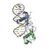
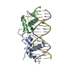
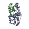
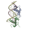
[English] 日本語
Yorodumi
- PDB-5e69: Glucocorticoid receptor DNA binding domain - IL8 NF-kB response e... -

+
Open data


ID or keywords:

Loading...

-
Basic information

Entry
Database: PDB / ID: 5.0E+69
TitleGlucocorticoid receptor DNA binding domain - IL8 NF-kB response element complex
Components
  • DNA (5'-D(*AP*TP*CP*GP*TP*GP*GP*AP*AP*TP*TP*TP*CP*CP*TP*C)-3')
  • DNA (5'-D(*GP*AP*GP*GP*AP*AP*AP*TP*TP*CP*CP*AP*CP*GP*AP*T)-3')
  • Glucocorticoid receptor
Keywordsdna binding protein/dna / DNA binding proteins / dna binding protein-dna complex
Function / homology
Function and homology information


Regulation of NPAS4 gene transcription / regulation of glucocorticoid biosynthetic process / nuclear glucocorticoid receptor activity / steroid hormone binding / PTK6 Expression / neuroinflammatory response / glucocorticoid metabolic process / microglia differentiation / mammary gland duct morphogenesis / maternal behavior ...Regulation of NPAS4 gene transcription / regulation of glucocorticoid biosynthetic process / nuclear glucocorticoid receptor activity / steroid hormone binding / PTK6 Expression / neuroinflammatory response / glucocorticoid metabolic process / microglia differentiation / mammary gland duct morphogenesis / maternal behavior / astrocyte differentiation / cellular response to glucocorticoid stimulus / motor behavior / adrenal gland development / regulation of gluconeogenesis / cellular response to steroid hormone stimulus / estrogen response element binding / FOXO-mediated transcription of oxidative stress, metabolic and neuronal genes / nuclear receptor-mediated steroid hormone signaling pathway / core promoter sequence-specific DNA binding / cellular response to transforming growth factor beta stimulus / HSP90 chaperone cycle for steroid hormone receptors (SHR) in the presence of ligand / steroid binding / TBP-class protein binding / cellular response to dexamethasone stimulus / synaptic transmission, glutamatergic / chromosome segregation / RNA polymerase II transcription regulatory region sequence-specific DNA binding / SUMOylation of intracellular receptors / Hsp90 protein binding / DNA-binding transcription repressor activity, RNA polymerase II-specific / positive regulation of miRNA transcription / Nuclear Receptor transcription pathway / spindle / Regulation of RUNX2 expression and activity / nuclear receptor activity / positive regulation of neuron apoptotic process / sequence-specific double-stranded DNA binding / Circadian Clock / chromatin organization / gene expression / DNA-binding transcription activator activity, RNA polymerase II-specific / Potential therapeutics for SARS / DNA-binding transcription factor activity, RNA polymerase II-specific / nuclear speck / mitochondrial matrix / RNA polymerase II cis-regulatory region sequence-specific DNA binding / DNA-binding transcription factor activity / cell division / negative regulation of DNA-templated transcription / centrosome / synapse / regulation of DNA-templated transcription / chromatin / regulation of transcription by RNA polymerase II / apoptotic process / protein kinase binding / negative regulation of transcription by RNA polymerase II / signal transduction / positive regulation of transcription by RNA polymerase II / protein-containing complex / RNA binding / zinc ion binding / nucleoplasm / identical protein binding / membrane / nucleus / cytosol / cytoplasm
Similarity search - Function
Glucocorticoid receptor / Glucocorticoid receptor / Erythroid Transcription Factor GATA-1, subunit A / Erythroid Transcription Factor GATA-1; Chain A / : / Nuclear hormone receptor / Nuclear hormones receptors DNA-binding region signature. / Zinc finger, nuclear hormone receptor-type / Zinc finger, C4 type (two domains) / Nuclear hormone receptors DNA-binding domain profile. ...Glucocorticoid receptor / Glucocorticoid receptor / Erythroid Transcription Factor GATA-1, subunit A / Erythroid Transcription Factor GATA-1; Chain A / : / Nuclear hormone receptor / Nuclear hormones receptors DNA-binding region signature. / Zinc finger, nuclear hormone receptor-type / Zinc finger, C4 type (two domains) / Nuclear hormone receptors DNA-binding domain profile. / c4 zinc finger in nuclear hormone receptors / Nuclear hormone receptor, ligand-binding domain / Nuclear hormone receptor-like domain superfamily / Ligand-binding domain of nuclear hormone receptor / Nuclear receptor (NR) ligand-binding (LBD) domain profile. / Ligand binding domain of hormone receptors / Zinc finger, NHR/GATA-type / 2-Layer Sandwich / Alpha Beta
Similarity search - Domain/homology
DNA / DNA (> 10) / Glucocorticoid receptor
Similarity search - Component
Biological speciesHomo sapiens (human)
MethodX-RAY DIFFRACTION / SYNCHROTRON / MOLECULAR REPLACEMENT / Resolution: 1.85 Å
AuthorsHudson, W.H. / Rye, E.A. / Herbst, A.G. / Ortlund, E.A.
CitationJournal: Nat Commun / Year: 2018
Title: Cryptic glucocorticoid receptor-binding sites pervade genomic NF-kappa B response elements.
Authors: Hudson, W.H. / Vera, I.M.S. / Nwachukwu, J.C. / Weikum, E.R. / Herbst, A.G. / Yang, Q. / Bain, D.L. / Nettles, K.W. / Kojetin, D.J. / Ortlund, E.A.
History
DepositionOct 9, 2015Deposition site: RCSB / Processing site: RCSB
Revision 1.0Feb 8, 2017Provider: repository / Type: Initial release
Revision 1.1Oct 24, 2018Group: Data collection / Database references / Category: citation / citation_author
Item: _citation.country / _citation.journal_abbrev ..._citation.country / _citation.journal_abbrev / _citation.journal_id_CSD / _citation.journal_id_ISSN / _citation.journal_volume / _citation.page_first / _citation.page_last / _citation.pdbx_database_id_DOI / _citation.pdbx_database_id_PubMed / _citation.title / _citation.year
Revision 1.2Mar 6, 2024Group: Data collection / Database references / Derived calculations
Category: chem_comp_atom / chem_comp_bond ...chem_comp_atom / chem_comp_bond / database_2 / struct_conn
Item: _database_2.pdbx_DOI / _database_2.pdbx_database_accession ..._database_2.pdbx_DOI / _database_2.pdbx_database_accession / _struct_conn.pdbx_dist_value / _struct_conn.ptnr1_auth_asym_id / _struct_conn.ptnr1_auth_comp_id / _struct_conn.ptnr1_auth_seq_id / _struct_conn.ptnr1_label_asym_id / _struct_conn.ptnr1_label_atom_id / _struct_conn.ptnr1_label_comp_id / _struct_conn.ptnr1_label_seq_id / _struct_conn.ptnr2_auth_asym_id / _struct_conn.ptnr2_auth_comp_id / _struct_conn.ptnr2_auth_seq_id / _struct_conn.ptnr2_label_asym_id / _struct_conn.ptnr2_label_atom_id / _struct_conn.ptnr2_label_comp_id

-
Structure visualization

Structure viewerMolecule:
MolmilJmol/JSmol

Downloads & links

-
Assembly

Deposited unit
A: Glucocorticoid receptor
C: DNA (5'-D(*GP*AP*GP*GP*AP*AP*AP*TP*TP*CP*CP*AP*CP*GP*AP*T)-3')
D: DNA (5'-D(*AP*TP*CP*GP*TP*GP*GP*AP*AP*TP*TP*TP*CP*CP*TP*C)-3')
B: Glucocorticoid receptor
hetero molecules


Theoretical massNumber of molelcules
Total (without water)35,5778
Polymers35,3154
Non-polymers2624
Water1,72996
1


  • Idetical with deposited unit
  • defined by author&software
TypeNameSymmetry operationNumber
identity operation1_555x,y,z1
Buried area4340 Å2
ΔGint-27 kcal/mol
Surface area12620 Å2
MethodPISA
Unit cell
Length a, b, c (Å)39.165, 97.176, 103.265
Angle α, β, γ (deg.)90.00, 90.00, 90.00
Int Tables number19
Space group name H-MP212121

-
Components

#1: Protein Glucocorticoid receptor / GR / Nuclear receptor subfamily 3 group C member 1


Mass: 12759.804 Da / Num. of mol.: 2 / Fragment: unp residues 391-480
Source method: isolated from a genetically manipulated source
Source: (gene. exp.) Homo sapiens (human) / Gene: NR3C1, GRL / Production host: Escherichia coli (E. coli) / Strain (production host): BL-21 (DE3) PLYSS / References: UniProt: P04150
#2: DNA chain DNA (5'-D(*GP*AP*GP*GP*AP*AP*AP*TP*TP*CP*CP*AP*CP*GP*AP*T)-3')


Mass: 4931.230 Da / Num. of mol.: 1 / Source method: obtained synthetically / Source: (synth.) Homo sapiens (human)
#3: DNA chain DNA (5'-D(*AP*TP*CP*GP*TP*GP*GP*AP*AP*TP*TP*TP*CP*CP*TP*C)-3')


Mass: 4864.164 Da / Num. of mol.: 1 / Source method: obtained synthetically / Source: (synth.) Homo sapiens (human)
#4: Chemical
ChemComp-ZN / ZINC ION


Mass: 65.409 Da / Num. of mol.: 4 / Source method: obtained synthetically / Formula: Zn
#5: Water ChemComp-HOH / water


Mass: 18.015 Da / Num. of mol.: 96 / Source method: isolated from a natural source / Formula: H2O

-
Experimental details

-
Experiment

ExperimentMethod: X-RAY DIFFRACTION

-
Sample preparation

CrystalDensity Matthews: 2.78 Å3/Da / Density % sol: 55.79 %
Crystal growTemperature: 298 K / Method: vapor diffusion, hanging drop / Details: 0.1 M HEPES pH 7.5, 7.5% glycerol, 22% PEG 20000

-
Data collection

DiffractionMean temperature: 100 K
Diffraction sourceSource: SYNCHROTRON / Site: APS / Beamline: 22-ID / Wavelength: 1 Å
DetectorType: MARMOSAIC 300 mm CCD / Detector: CCD / Date: Dec 12, 2012
RadiationProtocol: SINGLE WAVELENGTH / Monochromatic (M) / Laue (L): M / Scattering type: x-ray
Radiation wavelengthWavelength: 1 Å / Relative weight: 1
ReflectionResolution: 1.85→34.27 Å / Num. obs: 30412 / % possible obs: 88.4 % / Redundancy: 4.4 % / Net I/σ(I): 14
Reflection shellResolution: 1.85→1.92 Å / Redundancy: 2.3 % / Mean I/σ(I) obs: 2.8 / % possible all: 74.7

-
Processing

Software
NameVersionClassification
PHENIX1.9_1692refinement
SERGUIdata collection
HKL-2000data scaling
PHENIXphasing
RefinementMethod to determine structure: MOLECULAR REPLACEMENT / Resolution: 1.85→34.267 Å / SU ML: 0.23 / Cross valid method: FREE R-VALUE / σ(F): 1.34 / Phase error: 28.87 / Stereochemistry target values: ML
RfactorNum. reflection% reflectionSelection details
Rfree0.2418 1978 6.52 %Random selection
Rwork0.2197 ---
obs0.2212 30348 87.94 %-
Solvent computationShrinkage radii: 0.9 Å / VDW probe radii: 1.11 Å / Solvent model: FLAT BULK SOLVENT MODEL
Refinement stepCycle: LAST / Resolution: 1.85→34.267 Å
ProteinNucleic acidLigandSolventTotal
Num. atoms1120 650 4 96 1870
Refine LS restraints
Refine-IDTypeDev idealNumber
X-RAY DIFFRACTIONf_bond_d0.0071880
X-RAY DIFFRACTIONf_angle_d0.8522660
X-RAY DIFFRACTIONf_dihedral_angle_d21.94746
X-RAY DIFFRACTIONf_chiral_restr0.037288
X-RAY DIFFRACTIONf_plane_restr0.006228
LS refinement shell
Resolution (Å)Rfactor RfreeNum. reflection RfreeRfactor RworkNum. reflection RworkRefine-ID% reflection obs (%)
1.8502-1.89650.35121130.34311518X-RAY DIFFRACTION68
1.8965-1.94780.41951240.36581853X-RAY DIFFRACTION81
1.9478-2.00510.33711360.31391970X-RAY DIFFRACTION88
2.0051-2.06980.27321450.29852007X-RAY DIFFRACTION88
2.0698-2.14380.34191410.27292083X-RAY DIFFRACTION92
2.1438-2.22960.29341520.25982154X-RAY DIFFRACTION94
2.2296-2.3310.29561480.26632131X-RAY DIFFRACTION93
2.331-2.45390.28971520.24322158X-RAY DIFFRACTION94
2.4539-2.60760.28741490.24022113X-RAY DIFFRACTION93
2.6076-2.80880.25841550.23652107X-RAY DIFFRACTION92
2.8088-3.09130.23241390.23552107X-RAY DIFFRACTION91
3.0913-3.53830.24761390.21842090X-RAY DIFFRACTION89
3.5383-4.45630.21691480.19352067X-RAY DIFFRACTION88
4.4563-34.27320.18761370.17652012X-RAY DIFFRACTION81

+
About Yorodumi

-
News

-
Feb 9, 2022. New format data for meta-information of EMDB entries

New format data for meta-information of EMDB entries

  • Version 3 of the EMDB header file is now the official format.
  • The previous official version 1.9 will be removed from the archive.

Related info.:EMDB header

External links:wwPDB to switch to version 3 of the EMDB data model

-
Aug 12, 2020. Covid-19 info

Covid-19 info

URL: https://pdbjlc1.pdbj.org/emnavi/covid19.php

New page: Covid-19 featured information page in EM Navigator.

Related info.:Covid-19 info / Mar 5, 2020. Novel coronavirus structure data

+
Mar 5, 2020. Novel coronavirus structure data

Novel coronavirus structure data

Related info.:Yorodumi Speices / Aug 12, 2020. Covid-19 info

External links:COVID-19 featured content - PDBj / Molecule of the Month (242):Coronavirus Proteases

+
Jan 31, 2019. EMDB accession codes are about to change! (news from PDBe EMDB page)

EMDB accession codes are about to change! (news from PDBe EMDB page)

  • The allocation of 4 digits for EMDB accession codes will soon come to an end. Whilst these codes will remain in use, new EMDB accession codes will include an additional digit and will expand incrementally as the available range of codes is exhausted. The current 4-digit format prefixed with “EMD-” (i.e. EMD-XXXX) will advance to a 5-digit format (i.e. EMD-XXXXX), and so on. It is currently estimated that the 4-digit codes will be depleted around Spring 2019, at which point the 5-digit format will come into force.
  • The EM Navigator/Yorodumi systems omit the EMD- prefix.

Related info.:Q: What is EMD? / ID/Accession-code notation in Yorodumi/EM Navigator

External links:EMDB Accession Codes are Changing Soon! / Contact to PDBj

+
Jul 12, 2017. Major update of PDB

Major update of PDB

  • wwPDB released updated PDB data conforming to the new PDBx/mmCIF dictionary.
  • This is a major update changing the version number from 4 to 5, and with Remediation, in which all the entries are updated.
  • In this update, many items about electron microscopy experimental information are reorganized (e.g. em_software).
  • Now, EM Navigator and Yorodumi are based on the updated data.

External links:wwPDB Remediation / Enriched Model Files Conforming to OneDep Data Standards Now Available in the PDB FTP Archive

-
Yorodumi

Thousand views of thousand structures

  • Yorodumi is a browser for structure data from EMDB, PDB, SASBDB, etc.
  • This page is also the successor to EM Navigator detail page, and also detail information page/front-end page for Omokage search.
  • The word "yorodu" (or yorozu) is an old Japanese word meaning "ten thousand". "mi" (miru) is to see.

Related info.:EMDB / PDB / SASBDB / Comparison of 3 databanks / Yorodumi Search / Aug 31, 2016. New EM Navigator & Yorodumi / Yorodumi Papers / Jmol/JSmol / Function and homology information / Changes in new EM Navigator and Yorodumi

Read more