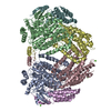
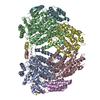
 Movie
Movie Controller


+ Open data

- Basic information
| Entry | Database: PDB / ID: 1mhy | |||||||||
|---|---|---|---|---|---|---|---|---|---|---|
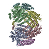
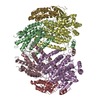
| Title | METHANE MONOOXYGENASE HYDROXYLASE | |||||||||
 Components Components | (METHANE MONOOXYGENASE HYDROXYLASE) x 3 | |||||||||
 Keywords Keywords | OXIDOREDUCTASE / MONOOXYGENASE / NADP / ONE-CARBON METABOLISM | |||||||||
| Function / homology |  Function and homology information Function and homology informationmethane metabolic process / methane monooxygenase (soluble) / methane monooxygenase [NAD(P)H] activity / one-carbon metabolic process / metal ion binding Similarity search - Function | |||||||||
| Biological species |  Methylosinus trichosporium (bacteria) Methylosinus trichosporium (bacteria) | |||||||||
| Method |  X-RAY DIFFRACTION / SYNCHROTRON / MOLECULAR REPLACEMENT / Resolution: 2 Å X-RAY DIFFRACTION / SYNCHROTRON / MOLECULAR REPLACEMENT / Resolution: 2 Å | |||||||||
 Authors Authors | Elango, N. / Radhakrishnan, R. / Froland, W.A. / Waller, B.J. / Earhart, C.A. / Lipscomb, J.D. / Ohlendorf, D.H. | |||||||||
 Citation Citation | Journal: Protein Sci. / Year: 1997 Title: Crystal structure of the hydroxylase component of methane monooxygenase from Methylosinus trichosporium OB3b Authors: Elango, N. / Radhakrishnan, R. / Froland, W.A. / Wallar, B.J. / Earhart, C.A. / Lipscomb, J.D. / Ohlendorf, D.H. | |||||||||
| History |
|
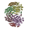
- Structure visualization
| Structure viewer | Molecule: MolmilJmol/JSmol |
|---|
- Downloads & links
-Download
| PDBx/mmCIF format | 1mhy.cif.gz | 310.1 KB | Display | PDBx/mmCIF format |
|---|---|---|---|---|
| PDB format | pdb1mhy.ent.gz | 247.9 KB | Display | PDB format |
| PDBx/mmJSON format | 1mhy.json.gz | Tree view | PDBx/mmJSON format | |
| Others |  Other downloads Other downloads |
-Validation report
| Arichive directory | https://data.pdbj.org/pub/pdb/validation_reports/mh/1mhyftp://data.pdbj.org/pub/pdb/validation_reports/mh/1mhy | HTTPS FTP |
|---|
-Related structure data
| Related structure data |  1mhzC  1mmoS S: Starting model for refinement C: citing same article ( |
|---|---|
| Similar structure data |


-Links
 PDBj
PDBj
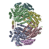
- Assembly
| Deposited unit | 
| ||||||||||||
|---|---|---|---|---|---|---|---|---|---|---|---|---|---|
| 1 | 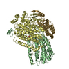
| ||||||||||||
| Unit cell |
| ||||||||||||
| Components on special symmetry positions |
|
-Components
| #1: Protein | Mass: 45165.145 Da / Num. of mol.: 1 / Source method: isolated from a natural source / Source: (natural) Methylosinus trichosporium (bacteria)References: UniProt: P27354, methane monooxygenase (soluble) | ||
|---|---|---|---|
| #2: Protein | Mass: 59513.691 Da / Num. of mol.: 1 / Source method: isolated from a natural source / Source: (natural) Methylosinus trichosporium (bacteria)References: UniProt: P27353, methane monooxygenase (soluble) | ||
| #3: Protein | Mass: 19358.285 Da / Num. of mol.: 1 / Source method: isolated from a natural source / Source: (natural) Methylosinus trichosporium (bacteria)References: UniProt: P27355, methane monooxygenase (soluble) | ||
| #4: Chemical |   Mass: 55.845 Da / Num. of mol.: 2 / Source method: obtained synthetically / Formula: Fe Mass: 55.845 Da / Num. of mol.: 2 / Source method: obtained synthetically / Formula: Fe#5: Water | ChemComp-HOH / |  Mass: 18.015 Da / Num. of mol.: 733 / Source method: isolated from a natural source / Formula: H2O Mass: 18.015 Da / Num. of mol.: 733 / Source method: isolated from a natural source / Formula: H2O |
-Experimental details
-Experiment
| Experiment | Method: X-RAY DIFFRACTION / Number of used crystals: 9 |
|---|
- Sample preparation
| Crystal | Density Matthews: 2.64 Å3/Da / Density % sol: 54 % | |||||||||||||||||||||||||
|---|---|---|---|---|---|---|---|---|---|---|---|---|---|---|---|---|---|---|---|---|---|---|---|---|---|---|
| Crystal grow | Details: FROLAND W.A. ET AL. (1994) J. MOL. BIOL. VOL. 236:379-381. | |||||||||||||||||||||||||
| Crystal grow | *PLUS Method: vapor diffusion, hanging dropDetails: used to seeding, Froland, W.A., (1994) J. Mol. Biol., 236, 379. PH range low: 7.8 / PH range high: 7.4 | |||||||||||||||||||||||||
| Components of the solutions | *PLUS
|
-Data collection
| Diffraction | Mean temperature: 291 K |
|---|---|
| Diffraction source | Source: SYNCHROTRON / Site: NSLS  / Beamline: X12C / Wavelength: 1.1 / Beamline: X12C / Wavelength: 1.1 |
| Detector | Type: MARRESEARCH / Detector: IMAGE PLATE / Date: May 10, 1994 |
| Radiation | Monochromatic (M) / Laue (L): M / Scattering type: x-ray |
| Radiation wavelength | Wavelength: 1.1 Å / Relative weight: 1 |
| Reflection | Num. obs: 87489 / % possible obs: 96.8 % / Observed criterion σ(I): 1 / Redundancy: 4 % / Rmerge(I) obs: 0.075 / Net I/σ(I): 12.5 |
| Reflection | *PLUS Num. measured all: 391667 |
- Processing
| Software |
| ||||||||||||||||||||||||||||||||||||||||||||||||||||||||||||
|---|---|---|---|---|---|---|---|---|---|---|---|---|---|---|---|---|---|---|---|---|---|---|---|---|---|---|---|---|---|---|---|---|---|---|---|---|---|---|---|---|---|---|---|---|---|---|---|---|---|---|---|---|---|---|---|---|---|---|---|---|---|
| Refinement | Method to determine structure: MOLECULAR REPLACEMENT Starting model: CHAINS B, D, AND G OF PDB ENTRY 1MMO Resolution: 2→5 Å / σ(F): 2
| ||||||||||||||||||||||||||||||||||||||||||||||||||||||||||||
| Displacement parameters | Biso mean: 23.9 Å2 | ||||||||||||||||||||||||||||||||||||||||||||||||||||||||||||
| Refine analyze | Luzzati coordinate error obs: 0.15 Å / Luzzati d res low obs: 5 Å | ||||||||||||||||||||||||||||||||||||||||||||||||||||||||||||
| Refinement step | Cycle: LAST / Resolution: 2→5 Å
| ||||||||||||||||||||||||||||||||||||||||||||||||||||||||||||
| Refine LS restraints |
| ||||||||||||||||||||||||||||||||||||||||||||||||||||||||||||
| Software | *PLUS Name: X-PLOR / Version: 3.1 / Classification: refinement | ||||||||||||||||||||||||||||||||||||||||||||||||||||||||||||
| Refinement | *PLUS Rfactor all: 0.145 | ||||||||||||||||||||||||||||||||||||||||||||||||||||||||||||
| Solvent computation | *PLUS | ||||||||||||||||||||||||||||||||||||||||||||||||||||||||||||
| Displacement parameters | *PLUS | ||||||||||||||||||||||||||||||||||||||||||||||||||||||||||||
| Refine LS restraints | *PLUS
|
