 Movie
Movie Controller


+ Open data

- Basic information
| Entry | Database: PDB / ID: 1mhx | ||||||
|---|---|---|---|---|---|---|---|
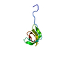
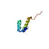
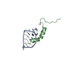
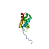
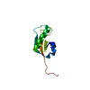
| Title | Crystal Structures of the redesigned protein G variant NuG1 | ||||||
 Components Components | immunoglobulin-binding protein G | ||||||
 Keywords Keywords | IMMUNE SYSTEM / alpha-beta protein / redesigned first beta-hairpin | ||||||
| Function / homology |  Function and homology information Function and homology informationProtein L, Ig light chain-binding / Protein L b1 domain / Ubiquitin-like (UB roll) - #10 / IgG-binding B / B domain / Ubiquitin-like (UB roll) / Roll / Alpha Beta Similarity search - Domain/homology | ||||||
| Biological species |  Finegoldia magna (bacteria) Finegoldia magna (bacteria) | ||||||
| Method |  X-RAY DIFFRACTION / SYNCHROTRON / MOLECULAR REPLACEMENT / Resolution: 1.8 Å X-RAY DIFFRACTION / SYNCHROTRON / MOLECULAR REPLACEMENT / Resolution: 1.8 Å | ||||||
 Authors Authors | Nauli, S. / Kuhlman, B. / Le Trong, I. / Stenkamp, R.E. / Teller, D.C. / Baker, D. | ||||||
 Citation Citation | Journal: Protein Sci. / Year: 2002 Title: Crystal structures and increased stabilization of the protein G variants with switched folding pathways NuG1 and NuG2 Authors: Nauli, S. / Kuhlman, B. / Le Trong, I. / Stenkamp, R.E. / Teller, D.C. / Baker, D. | ||||||
| History |
| ||||||
| Remark 999 | SEQUENCE THE SEQUENCE DIFFERS FROM PIR ENTRY A45063 AT RESIDUES 15-25 (PIR RESIDUES 334-344) ...SEQUENCE THE SEQUENCE DIFFERS FROM PIR ENTRY A45063 AT RESIDUES 15-25 (PIR RESIDUES 334-344) BECAUSE THE AUTHORS REDESIGNED THE FIRST HAIRPIN. |
- Structure visualization
| Structure viewer | Molecule: MolmilJmol/JSmol |
|---|
- Downloads & links
-Download
| PDBx/mmCIF format | 1mhx.cif.gz | 26.2 KB | Display | PDBx/mmCIF format |
|---|---|---|---|---|
| PDB format | pdb1mhx.ent.gz | 16.9 KB | Display | PDB format |
| PDBx/mmJSON format | 1mhx.json.gz | Tree view | PDBx/mmJSON format | |
| Others |  Other downloads Other downloads |
-Validation report
| Arichive directory | https://data.pdbj.org/pub/pdb/validation_reports/mh/1mhxftp://data.pdbj.org/pub/pdb/validation_reports/mh/1mhx | HTTPS FTP |
|---|
-Related structure data
| Related structure data | 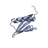 1mi0C 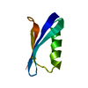 1pgaS C: citing same article ( S: Starting model for refinement |
|---|---|
| Similar structure data |

-Links
 PDBj
PDBj- Assembly
| Deposited unit | 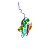
| ||||||||
|---|---|---|---|---|---|---|---|---|---|
| 1 |
| ||||||||
| Unit cell |
| ||||||||
| Components on special symmetry positions |
|
-Components
| #1: Protein | Mass: 7359.115 Da / Num. of mol.: 1 / Fragment: Redesigned B1 domain / Mutation: T58A Source method: isolated from a genetically manipulated source Details: Redesigned first beta-hairpin, variant NuG1 / Source: (gene. exp.) Finegoldia magna (bacteria) / Strain: ATCC 29328 / Production host: |
|---|---|
| #2: Water | ChemComp-HOH /  Mass: 18.015 Da / Num. of mol.: 87 / Source method: isolated from a natural source / Formula: H2O Mass: 18.015 Da / Num. of mol.: 87 / Source method: isolated from a natural source / Formula: H2O |
-Experimental details
-Experiment
| Experiment | Method: X-RAY DIFFRACTION / Number of used crystals: 1 |
|---|
- Sample preparation
| Crystal | Density Matthews: 2.14 Å3/Da / Density % sol: 42.53 % | ||||||||||||||||||||||||
|---|---|---|---|---|---|---|---|---|---|---|---|---|---|---|---|---|---|---|---|---|---|---|---|---|---|
| Crystal grow | Temperature: 295 K / Method: vapor diffusion, sitting drop / pH: 7.5 Details: n-propanol, sodium formate, tris-hcl, pH 7.5, VAPOR DIFFUSION, SITTING DROP, temperature 295K | ||||||||||||||||||||||||
| Crystal grow | *PLUS Method: vapor diffusion, hanging drop | ||||||||||||||||||||||||
| Components of the solutions | *PLUS
|
-Data collection
| Diffraction | Mean temperature: 298 K |
|---|---|
| Diffraction source | Source: SYNCHROTRON / Site: SSRL  / Beamline: BL9-1 / Wavelength: 1 Å / Beamline: BL9-1 / Wavelength: 1 Å |
| Detector | Type: ADSC QUANTUM 4 / Detector: CCD / Date: Apr 20, 2001 |
| Radiation | Monochromator: graphite / Protocol: SINGLE WAVELENGTH / Monochromatic (M) / Laue (L): M / Scattering type: x-ray |
| Radiation wavelength | Wavelength: 1 Å / Relative weight: 1 |
| Reflection | Resolution: 1.8→22.3 Å / Num. obs: 6273 / % possible obs: 95.4 % / Observed criterion σ(F): 2 / Observed criterion σ(I): 2.2 |
| Reflection shell | Resolution: 1.8→1.86 Å / % possible all: 92.5 |
| Reflection | *PLUS Num. obs: 6383 / Rmerge(I) obs: 0.116 |
- Processing
| Software |
| ||||||||||||||||||||
|---|---|---|---|---|---|---|---|---|---|---|---|---|---|---|---|---|---|---|---|---|---|
| Refinement | Method to determine structure: MOLECULAR REPLACEMENT Starting model: pdb entry 1pga Resolution: 1.8→22.3 Å / σ(F): 2
| ||||||||||||||||||||
| Refinement step | Cycle: LAST / Resolution: 1.8→22.3 Å
| ||||||||||||||||||||
| Refine LS restraints |
| ||||||||||||||||||||
| Refine LS restraints | *PLUS Type: x_bond_d / Dev ideal: 0.006 |
