 Movie
Movie Controller


+ Open data

- Basic information
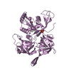
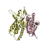
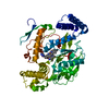
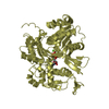
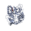
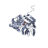
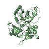
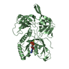
| Entry | Database: PDB / ID: 1mh3 | ||||||
|---|---|---|---|---|---|---|---|
| Title | maltose binding-a1 homeodomain protein chimera, crystal form I | ||||||
 Components Components | maltose binding-a1 homeodomain protein chimera | ||||||
 Keywords Keywords | SUGAR BINDING / DNA BINDING PROTEIN / MATa1 / homeodomain / binding cooperativity / maltose binding protein / MBP | ||||||
| Function / homology |  Function and homology information Function and homology informationdetection of maltose stimulus / maltose transport complex / carbohydrate transport / carbohydrate transmembrane transporter activity / maltose binding / maltose transport / maltodextrin transmembrane transport / ATP-binding cassette (ABC) transporter complex, substrate-binding subunit-containing / ATP-binding cassette (ABC) transporter complex / cell chemotaxis ...detection of maltose stimulus / maltose transport complex / carbohydrate transport / carbohydrate transmembrane transporter activity / maltose binding / maltose transport / maltodextrin transmembrane transport / ATP-binding cassette (ABC) transporter complex, substrate-binding subunit-containing / ATP-binding cassette (ABC) transporter complex / cell chemotaxis / outer membrane-bounded periplasmic space / DNA-binding transcription factor activity, RNA polymerase II-specific / periplasmic space / DNA damage response / DNA binding / membrane / nucleus Similarity search - Function | ||||||
| Biological species |   | ||||||
| Method |  X-RAY DIFFRACTION / MOLECULAR REPLACEMENT / Resolution: 2.1 Å X-RAY DIFFRACTION / MOLECULAR REPLACEMENT / Resolution: 2.1 Å | ||||||
 Authors Authors | Ke, A. / Wolberger, C. | ||||||
 Citation Citation | Journal: Protein Sci. / Year: 2003 Title: Insights into binding cooperativity of MATa1/MATalpha2 from the crystal structure of a MATa1 homeodomain-maltose binding protein chimera Authors: Ke, A. / Wolberger, C. | ||||||
| History |
| ||||||
| Remark 999 | SEQUENCE THERE IS A 5 ALANINE LINKER WHICH LINKS THE MALTOSE-BINDING DOMAIN TO THE MATA1 HOMEODOMAIN. |
- Structure visualization
| Structure viewer | Molecule: MolmilJmol/JSmol |
|---|
- Downloads & links
-Download
| PDBx/mmCIF format | 1mh3.cif.gz | 96.1 KB | Display | PDBx/mmCIF format |
|---|---|---|---|---|
| PDB format | pdb1mh3.ent.gz | 73.4 KB | Display | PDB format |
| PDBx/mmJSON format | 1mh3.json.gz | Tree view | PDBx/mmJSON format | |
| Others |  Other downloads Other downloads |
-Validation report
| Arichive directory | https://data.pdbj.org/pub/pdb/validation_reports/mh/1mh3ftp://data.pdbj.org/pub/pdb/validation_reports/mh/1mh3 | HTTPS FTP |
|---|
-Related structure data
| Related structure data | 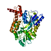 1mh4C  4mbpS C: citing same article ( S: Starting model for refinement |
|---|---|
| Similar structure data |

-Links
 PDBj
PDBj


- Assembly
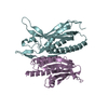
| Deposited unit | 
| ||||||||
|---|---|---|---|---|---|---|---|---|---|
| 1 |
| ||||||||
| Unit cell |
|
-Components
| #1: Protein | Mass: 46403.832 Da / Num. of mol.: 1 / Mutation: E359A, K362A, D363A Source method: isolated from a genetically manipulated source Details: Chimera consisting of residues 1-366 (sequence database residues 27-392) of Maltose-binding periplasmic protein, a 5 alanine linker, and residues 477-526 (sequence database residues 77-126) ...Details: Chimera consisting of residues 1-366 (sequence database residues 27-392) of Maltose-binding periplasmic protein, a 5 alanine linker, and residues 477-526 (sequence database residues 77-126) of homeobox of mating-type protein A-1. Source: (gene. exp.) Genus: Escherichia, Saccharomyces / Species: , / Strain: , / Plasmid: pMAL-c2 with modifications at the fusion linker / Production host: References: UniProt: P02928, UniProt: P01366, UniProt: P0AEX9*PLUS |
|---|---|
| #2: Water | ChemComp-HOH /  Mass: 18.015 Da / Num. of mol.: 230 / Source method: isolated from a natural source / Formula: H2O Mass: 18.015 Da / Num. of mol.: 230 / Source method: isolated from a natural source / Formula: H2O |
-Experimental details
-Experiment
| Experiment | Method: X-RAY DIFFRACTION / Number of used crystals: 1 |
|---|
- Sample preparation
| Crystal | Density Matthews: 3.83 Å3/Da / Density % sol: 67.86 % | ||||||||||||||||||||||||
|---|---|---|---|---|---|---|---|---|---|---|---|---|---|---|---|---|---|---|---|---|---|---|---|---|---|
| Crystal grow | Temperature: 298 K / Method: vapor diffusion, hanging drop / pH: 5 Details: amonium sulfate, pH 5.0, VAPOR DIFFUSION, HANGING DROP, temperature 298K | ||||||||||||||||||||||||
| Crystal grow | *PLUS | ||||||||||||||||||||||||
| Components of the solutions | *PLUS
|
-Data collection
| Diffraction | Mean temperature: 100 K |
|---|---|
| Diffraction source | Source: ROTATING ANODE / Type: RIGAKU RU200 / Wavelength: 1.5418 |
| Detector | Type: RIGAKU RAXIS IV / Detector: IMAGE PLATE |
| Radiation | Monochromator: YALE MIRRORS / Protocol: SINGLE WAVELENGTH / Monochromatic (M) / Laue (L): M / Scattering type: x-ray |
| Radiation wavelength | Wavelength: 1.5418 Å / Relative weight: 1 |
| Reflection | Resolution: 2.1→45.64 Å / Num. all: 42955 / Num. obs: 42612 / % possible obs: 99.2 % / Observed criterion σ(F): 3 / Observed criterion σ(I): 4.9 / Redundancy: 6.8 % / Biso Wilson estimate: 17.9 Å2 / Rsym value: 0.057 / Net I/σ(I): 30.1 |
| Reflection shell | Resolution: 2.1→2.2 Å / Mean I/σ(I) obs: 4.9 / Rsym value: 0.216 / % possible all: 97.1 |
| Reflection | *PLUS Lowest resolution: 60 Å / Rmerge(I) obs: 0.057 |
| Reflection shell | *PLUS % possible obs: 97.1 % / Rmerge(I) obs: 0.216 |
- Processing
| Software |
| |||||||||||||||||||||||||
|---|---|---|---|---|---|---|---|---|---|---|---|---|---|---|---|---|---|---|---|---|---|---|---|---|---|---|
| Refinement | Method to determine structure: MOLECULAR REPLACEMENT Starting model: MBP (pdb code 4MBP) Resolution: 2.1→45.64 Å / Rfactor Rfree error: 0.006 / Isotropic thermal model: RESTRAINED / Cross valid method: THROUGHOUT / σ(F): 4.9 / σ(I): 4.9 / Stereochemistry target values: Engh & Huber
| |||||||||||||||||||||||||
| Solvent computation | Solvent model: FLAT MODEL / Bsol: 49.3184 Å2 / ksol: 0.377928 e/Å3 | |||||||||||||||||||||||||
| Displacement parameters | Biso mean: 24.9 Å2
| |||||||||||||||||||||||||
| Refine analyze | Luzzati coordinate error free: 0.31 Å / Luzzati sigma a free: 0.2 Å | |||||||||||||||||||||||||
| Refinement step | Cycle: LAST / Resolution: 2.1→45.64 Å
| |||||||||||||||||||||||||
| Refine LS restraints |
| |||||||||||||||||||||||||
| LS refinement shell | Highest resolution: 2.1 Å / Total num. of bins used: 6
| |||||||||||||||||||||||||
| Xplor file |
| |||||||||||||||||||||||||
| Refinement | *PLUS Highest resolution: 2.1 Å / Lowest resolution: 60 Å / Rfactor Rwork: 0.231 | |||||||||||||||||||||||||
| Solvent computation | *PLUS | |||||||||||||||||||||||||
| Displacement parameters | *PLUS | |||||||||||||||||||||||||
| Refine LS restraints | *PLUS
|
