 Movie
Movie Controller


+ Open data

- Basic information
| Entry | Database: PDB / ID: 1mg1 | |||||||||
|---|---|---|---|---|---|---|---|---|---|---|
| Title | HTLV-1 GP21 ECTODOMAIN/MALTOSE-BINDING PROTEIN CHIMERA | |||||||||
 Components Components | PROTEIN (HTLV-1 GP21 ECTODOMAIN/MALTOSE-BINDING PROTEIN CHIMERA) | |||||||||
 Keywords Keywords | VIRAL PROTEIN / HUMAN T CELL LEUKEMIA VIRUS TYPE 1 / HTLV-1 / ENVELOPE PROTEIN / MEMBRANE FUSION / MALTOSE-BINDING PROTEIN CHIMERA | |||||||||
| Function / homology |  Function and homology information Function and homology informationdetection of maltose stimulus / maltose transport complex / carbohydrate transport / carbohydrate transmembrane transporter activity / maltose binding / maltose transport / maltodextrin transmembrane transport / ATP-binding cassette (ABC) transporter complex, substrate-binding subunit-containing / ATP-binding cassette (ABC) transporter complex / cell chemotaxis ...detection of maltose stimulus / maltose transport complex / carbohydrate transport / carbohydrate transmembrane transporter activity / maltose binding / maltose transport / maltodextrin transmembrane transport / ATP-binding cassette (ABC) transporter complex, substrate-binding subunit-containing / ATP-binding cassette (ABC) transporter complex / cell chemotaxis / outer membrane-bounded periplasmic space / periplasmic space / DNA damage response / membrane Similarity search - Function | |||||||||
| Biological species |  Human T-lymphotropic virus 1 Human T-lymphotropic virus 1 | |||||||||
| Method |  X-RAY DIFFRACTION / MOLECULAR REPLACEMENT / Resolution: 2.5 Å X-RAY DIFFRACTION / MOLECULAR REPLACEMENT / Resolution: 2.5 Å | |||||||||
 Authors Authors | Kobe, B. / Center, R.J. / Kemp, B.E. / Poumbourios, P. | |||||||||
 Citation Citation | Journal: Proc.Natl.Acad.Sci.USA / Year: 1999 Title: Crystal structure of human T cell leukemia virus type 1 gp21 ectodomain crystallized as a maltose-binding protein chimera reveals structural evolution of retroviral transmembrane proteins. Authors: Kobe, B. / Center, R.J. / Kemp, B.E. / Poumbourios, P. #1: Journal: Protein Sci. / Year: 1998Title: Crystallization of a Trimeric Human T Cell Leukemia Virus Type 1 Gp21 Ectodomain Fragment as a Chimera with Maltose-Binding Protein Authors: Center, R.J. / Kobe, B. / Wilson, K.A. / Teh, T. / Howlett, G.J. / Kemp, B.E. / Poumbourios, P. | |||||||||
| History |
|
- Structure visualization
| Structure viewer | Molecule: MolmilJmol/JSmol |
|---|
- Downloads & links
-Download
| PDBx/mmCIF format | 1mg1.cif.gz | 101.5 KB | Display | PDBx/mmCIF format |
|---|---|---|---|---|
| PDB format | pdb1mg1.ent.gz | 76.3 KB | Display | PDB format |
| PDBx/mmJSON format | 1mg1.json.gz | Tree view | PDBx/mmJSON format | |
| Others |  Other downloads Other downloads |
-Validation report
| Arichive directory | https://data.pdbj.org/pub/pdb/validation_reports/mg/1mg1ftp://data.pdbj.org/pub/pdb/validation_reports/mg/1mg1 | HTTPS FTP |
|---|
-Related structure data
| Related structure data |  1anfS S: Starting model for refinement |
|---|---|
| Similar structure data |


-Links
 PDBj
PDBj


- Assembly
| Deposited unit | 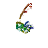
| ||||||||||
|---|---|---|---|---|---|---|---|---|---|---|---|
| 1 | 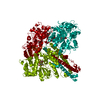
| ||||||||||
| Unit cell |
| ||||||||||
| Components on special symmetry positions |
|
-Components
| #1: Protein | Mass: 49386.023 Da / Num. of mol.: 1 / Fragment: GP21 ECTODOMAIN (GP62 RESIDUES 338-425) Source method: isolated from a genetically manipulated source Source: (gene. exp.) Human T-lymphotropic virus 1 / Genus: Deltaretrovirus / Species: Primate T-lymphotropic virus 1 / Strain: 3-19-3 / Cellular location: VIRUS ENVELOPE / Production host:  | ||||
|---|---|---|---|---|---|
| #2: Polysaccharide | alpha-D-glucopyranose-(1-4)-alpha-D-glucopyranose / alpha-maltose  Source method: isolated from a genetically manipulated source Details: oligosaccharide / References: alpha-maltose | ||||
| #3: Chemical |   Mass: 35.453 Da / Num. of mol.: 2 / Source method: obtained synthetically / Formula: Cl Mass: 35.453 Da / Num. of mol.: 2 / Source method: obtained synthetically / Formula: Cl#4: Water | ChemComp-HOH / |  Mass: 18.015 Da / Num. of mol.: 37 / Source method: isolated from a natural source / Formula: H2O Mass: 18.015 Da / Num. of mol.: 37 / Source method: isolated from a natural source / Formula: H2OHas protein modification | Y | |
-Experimental details
-Experiment
| Experiment | Method: X-RAY DIFFRACTION / Number of used crystals: 1 |
|---|
- Sample preparation
| Crystal | Density Matthews: 2.34 Å3/Da / Density % sol: 47 % | ||||||||||||||||||||
|---|---|---|---|---|---|---|---|---|---|---|---|---|---|---|---|---|---|---|---|---|---|
| Crystal grow | pH: 4.7 / Details: pH 4.7 | ||||||||||||||||||||
| Components of the solutions |
| ||||||||||||||||||||
| Crystal grow | *PLUS Method: vapor diffusion, hanging drop | ||||||||||||||||||||
| Components of the solutions | *PLUS
|
-Data collection
| Diffraction | Mean temperature: 100 K |
|---|---|
| Diffraction source | Source: ROTATING ANODE / Type: RIGAKU RU200 / Wavelength: 1.5418 |
| Detector | Type: MARRESEARCH / Detector: IMAGE PLATE / Date: Apr 15, 1998 |
| Radiation | Monochromator: GRAPHITE / Protocol: SINGLE WAVELENGTH / Monochromatic (M) / Laue (L): M / Scattering type: x-ray |
| Radiation wavelength | Wavelength: 1.5418 Å / Relative weight: 1 |
| Reflection | Resolution: 2.29→99 Å / Num. obs: 19248 / % possible obs: 91 % / Redundancy: 7.8 % / Biso Wilson estimate: 60.4 Å2 / Rmerge(I) obs: 0.068 / Net I/σ(I): 9.8 |
| Reflection shell | Resolution: 2.29→2.37 Å / Rmerge(I) obs: 0.364 / Mean I/σ(I) obs: 1.2 / % possible all: 65 |
| Reflection | *PLUS Num. measured all: 150336 |
| Reflection shell | *PLUS % possible obs: 65 % |
- Processing
| Software |
| ||||||||||||||||||||||||||||||||||||||||||||||||||||||||||||||||||||||||||||||||
|---|---|---|---|---|---|---|---|---|---|---|---|---|---|---|---|---|---|---|---|---|---|---|---|---|---|---|---|---|---|---|---|---|---|---|---|---|---|---|---|---|---|---|---|---|---|---|---|---|---|---|---|---|---|---|---|---|---|---|---|---|---|---|---|---|---|---|---|---|---|---|---|---|---|---|---|---|---|---|---|---|---|
| Refinement | Method to determine structure: MOLECULAR REPLACEMENT Starting model: 1ANF Resolution: 2.5→40 Å / Rfactor Rfree error: 0.009 / Data cutoff high absF: 1000000 / Data cutoff low absF: 0.001 / Isotropic thermal model: RESTRAINED / Cross valid method: THROUGHOUT / σ(F): 0 / Details: BULK SOLVENT MODEL USED
| ||||||||||||||||||||||||||||||||||||||||||||||||||||||||||||||||||||||||||||||||
| Displacement parameters | Biso mean: 65.3 Å2
| ||||||||||||||||||||||||||||||||||||||||||||||||||||||||||||||||||||||||||||||||
| Refine analyze |
| ||||||||||||||||||||||||||||||||||||||||||||||||||||||||||||||||||||||||||||||||
| Refinement step | Cycle: LAST / Resolution: 2.5→40 Å
| ||||||||||||||||||||||||||||||||||||||||||||||||||||||||||||||||||||||||||||||||
| Refine LS restraints |
| ||||||||||||||||||||||||||||||||||||||||||||||||||||||||||||||||||||||||||||||||
| LS refinement shell | Resolution: 2.5→2.59 Å / Rfactor Rfree error: 0.044 / Total num. of bins used: 10
| ||||||||||||||||||||||||||||||||||||||||||||||||||||||||||||||||||||||||||||||||
| Xplor file |
| ||||||||||||||||||||||||||||||||||||||||||||||||||||||||||||||||||||||||||||||||
| Software | *PLUS Name: X-PLOR / Version: 3.851 / Classification: refinement | ||||||||||||||||||||||||||||||||||||||||||||||||||||||||||||||||||||||||||||||||
| Refinement | *PLUS Rfactor obs: 0.223 | ||||||||||||||||||||||||||||||||||||||||||||||||||||||||||||||||||||||||||||||||
| Solvent computation | *PLUS | ||||||||||||||||||||||||||||||||||||||||||||||||||||||||||||||||||||||||||||||||
| Displacement parameters | *PLUS | ||||||||||||||||||||||||||||||||||||||||||||||||||||||||||||||||||||||||||||||||
| Refine LS restraints | *PLUS
| ||||||||||||||||||||||||||||||||||||||||||||||||||||||||||||||||||||||||||||||||
| LS refinement shell | *PLUS Rfactor obs: 0.44 |
