 Movie
Movie Controller

[English] 日本語
 Yorodumi
- PDB-1mfu: Probing the role of a mobile loop in human salivary amylase: Stru... -
+ Open data

- Basic information
| Entry | Database: PDB / ID: 1mfu | ||||||||||||
|---|---|---|---|---|---|---|---|---|---|---|---|---|---|
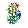
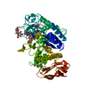
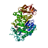
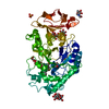
| Title | Probing the role of a mobile loop in human salivary amylase: Structural studies on the loop-deleted mutant | ||||||||||||
 Components Components | alpha-amylase, Salivary | ||||||||||||
 Keywords Keywords | HYDROLASE / amylase / acarbose complex / mutageneis / mobile loop / deletion mutant | ||||||||||||
| Function / homology |  Function and homology information Function and homology informationDigestion of dietary carbohydrate / oligosaccharide metabolic process / alpha-amylase / alpha-amylase activity / chloride ion binding / carbohydrate metabolic process / calcium ion binding / : / extracellular exosome Similarity search - Function | ||||||||||||
| Biological species |  Homo sapiens (human) Homo sapiens (human) | ||||||||||||
| Method |  X-RAY DIFFRACTION / SYNCHROTRON / MOLECULAR REPLACEMENT / Resolution: 2 Å X-RAY DIFFRACTION / SYNCHROTRON / MOLECULAR REPLACEMENT / Resolution: 2 Å | ||||||||||||
 Authors Authors | Ramasubbu, N. / Ragunath, C. / Mishra, P.J. | ||||||||||||
 Citation Citation | Journal: J.Mol.Biol. / Year: 2003 Title: Probing the role of a mobile loop in substrate binding and enzyme activity of human salivary amylase. Authors: Ramasubbu, N. / Ragunath, C. / Mishra, P.J. | ||||||||||||
| History |
|
- Structure visualization
| Structure viewer | Molecule: MolmilJmol/JSmol |
|---|
- Downloads & links
-Download
| PDBx/mmCIF format | 1mfu.cif.gz | 123.9 KB | Display | PDBx/mmCIF format |
|---|---|---|---|---|
| PDB format | pdb1mfu.ent.gz | 94.8 KB | Display | PDB format |
| PDBx/mmJSON format | 1mfu.json.gz | Tree view | PDBx/mmJSON format | |
| Others |  Other downloads Other downloads |
-Validation report
| Arichive directory | https://data.pdbj.org/pub/pdb/validation_reports/mf/1mfuftp://data.pdbj.org/pub/pdb/validation_reports/mf/1mfu | HTTPS FTP |
|---|
-Related structure data
| Related structure data |  1jxkSC  1mfvC S: Starting model for refinement C: citing same article ( |
|---|---|
| Similar structure data |


-Links
 PDBj
PDBj


- Assembly
| Deposited unit | 
| ||||||||
|---|---|---|---|---|---|---|---|---|---|
| 1 |
| ||||||||
| Unit cell |
|
-Components
-Protein , 1 types, 1 molecules A
| #1: Protein | Mass: 55641.945 Da / Num. of mol.: 1 / Mutation: deletion of residues 306 through 310 Source method: isolated from a genetically manipulated source Source: (gene. exp.) Homo sapiens (human) / Plasmid: pVL1392 / Production host:   Spodoptera frugiperda (fall armyworm) Spodoptera frugiperda (fall armyworm)References: UniProt: P04745, UniProt: P0DTE8*PLUS, alpha-amylase |
|---|
-Sugars , 4 types, 6 molecules 
| #2: Polysaccharide | 4-amino-4,6-dideoxy-alpha-D-glucopyranose-(1-4)-alpha-D-glucopyranose Source method: isolated from a genetically manipulated source | ||||
|---|---|---|---|---|---|
| #3: Polysaccharide | Source method: isolated from a genetically manipulated source #4: Polysaccharide | alpha-D-glucopyranose-(1-4)-alpha-D-glucopyranose / alpha-maltose |   Source method: isolated from a genetically manipulated source Details: oligosaccharide / References: alpha-maltose #6: Sugar | ChemComp-GLC / |  Type: D-saccharide, alpha linking / Mass: 180.156 Da / Num. of mol.: 1 Type: D-saccharide, alpha linking / Mass: 180.156 Da / Num. of mol.: 1Source method: isolated from a genetically manipulated source Formula: C6H12O6 |
-Non-polymers , 4 types, 296 molecules 


| #5: Chemical | ChemComp-HMC /  Mass: 176.167 Da / Num. of mol.: 4 Mass: 176.167 Da / Num. of mol.: 4Source method: isolated from a genetically manipulated source Formula: C7H12O5 #7: Chemical | ChemComp-CA / |  Mass: 40.078 Da / Num. of mol.: 1 / Source method: obtained synthetically / Formula: Ca Mass: 40.078 Da / Num. of mol.: 1 / Source method: obtained synthetically / Formula: Ca#8: Chemical | ChemComp-CL / |  Mass: 35.453 Da / Num. of mol.: 1 / Source method: obtained synthetically / Formula: Cl Mass: 35.453 Da / Num. of mol.: 1 / Source method: obtained synthetically / Formula: Cl#9: Water | ChemComp-HOH / | Mass: 18.015 Da / Num. of mol.: 290 / Source method: isolated from a natural source / Formula: H2O |
|---|
-Details
| Has protein modification | Y |
|---|
-Experimental details
-Experiment
| Experiment | Method: X-RAY DIFFRACTION / Number of used crystals: 1 |
|---|
- Sample preparation
| Crystal | Density Matthews: 2.34 Å3/Da / Density % sol: 47.53 % |
|---|---|
| Crystal grow | Temperature: 298 K Method: soaking with acarbose at 1 mm concentration for 24 hours pH: 9 Details: 40% mpd, pH 9.0, soaking with acarbose at 1 mM concentration for 24 hours, temperature 298K |
-Data collection
| Diffraction | Mean temperature: 100 K |
|---|---|
| Diffraction source | Source: SYNCHROTRON / Site: CHESS  / Beamline: F1 / Wavelength: 0.91 Å / Beamline: F1 / Wavelength: 0.91 Å |
| Detector | Type: ADSC QUANTUM 4 / Detector: CCD / Date: Mar 31, 2002 / Details: monochromator |
| Radiation | Protocol: SINGLE WAVELENGTH / Monochromatic (M) / Laue (L): M / Scattering type: x-ray |
| Radiation wavelength | Wavelength: 0.91 Å / Relative weight: 1 |
| Reflection | Resolution: 2→26.54 Å / Num. all: 35031 / Num. obs: 35031 / % possible obs: 96.8 % / Observed criterion σ(F): 1 / Observed criterion σ(I): 1 / Redundancy: 4.8 % / Biso Wilson estimate: 25.6 Å2 / Rmerge(I) obs: 0.06 / Rsym value: 0.06 / Net I/σ(I): 24.5 |
| Reflection shell | Resolution: 2→2.07 Å / Rmerge(I) obs: 0.267 / Mean I/σ(I) obs: 7 / Num. unique all: 3488 / Rsym value: 0.267 |
- Processing
| Software |
| |||||||||||||||||||||||||||||||||||||||||||||||||||||||||||||||||||||||||||
|---|---|---|---|---|---|---|---|---|---|---|---|---|---|---|---|---|---|---|---|---|---|---|---|---|---|---|---|---|---|---|---|---|---|---|---|---|---|---|---|---|---|---|---|---|---|---|---|---|---|---|---|---|---|---|---|---|---|---|---|---|---|---|---|---|---|---|---|---|---|---|---|---|---|---|---|---|
| Refinement | Method to determine structure: MOLECULAR REPLACEMENT Starting model: PDB entry 1jxk Resolution: 2→26.54 Å / Cor.coef. Fo:Fc: 0.964 / Cor.coef. Fo:Fc free: 0.943 / Cross valid method: THROUGHOUT / σ(F): 0 / Stereochemistry target values: MAXIMUM LIKELIHOOD
| |||||||||||||||||||||||||||||||||||||||||||||||||||||||||||||||||||||||||||
| Solvent computation | Ion probe radii: 0.8 Å / Shrinkage radii: 0.8 Å / VDW probe radii: 1.4 Å / Solvent model: BABINET MODEL WITH MASK | |||||||||||||||||||||||||||||||||||||||||||||||||||||||||||||||||||||||||||
| Displacement parameters | Biso mean: 25.789 Å2
| |||||||||||||||||||||||||||||||||||||||||||||||||||||||||||||||||||||||||||
| Refine analyze |
| |||||||||||||||||||||||||||||||||||||||||||||||||||||||||||||||||||||||||||
| Refinement step | Cycle: LAST / Resolution: 2→26.54 Å
| |||||||||||||||||||||||||||||||||||||||||||||||||||||||||||||||||||||||||||
| Refine LS restraints |
| |||||||||||||||||||||||||||||||||||||||||||||||||||||||||||||||||||||||||||
| LS refinement shell | Resolution: 2.001→2.052 Å / Total num. of bins used: 20 /
|
