 Movie
Movie Controller


+ Open data

- Basic information
| Entry | Database: PDB / ID: 1mf5 | ||||||||||||||||||
|---|---|---|---|---|---|---|---|---|---|---|---|---|---|---|---|---|---|---|---|
| Title | GCATGCT Quadruplex | ||||||||||||||||||
 Components Components | 5'-D(* Keywords KeywordsDNA / QUADRUPLEX / ATOMIC RESOLUTION | Function / homology | COBALT HEXAMMINE(III) / DNA |  Function and homology information Function and homology informationMethod |  X-RAY DIFFRACTION / SYNCHROTRON / MOLECULAR REPLACEMENT / Resolution: 1.1 Å X-RAY DIFFRACTION / SYNCHROTRON / MOLECULAR REPLACEMENT / Resolution: 1.1 Å  Authors AuthorsThorpe, J.H. / Teixeira, S.C.M. / Gale, B.C. / Cardin, C.J. |  CitationJournal: Nucleic Acids Res. / Year: 2003 CitationJournal: Nucleic Acids Res. / Year: 2003Title: Crystal structure of the complementary quadruplex formed by d(GCATGCT) at atomic resolution Authors: Thorpe, J.H. / Teixeira, S.C.M. / Gale, B.C. / Cardin, C.J. History |
|
- Structure visualization
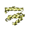
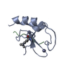
| Structure viewer | Molecule: MolmilJmol/JSmol |
|---|
- Downloads & links
-Download
| PDBx/mmCIF format | 1mf5.cif.gz | 25.9 KB | Display | PDBx/mmCIF format |
|---|---|---|---|---|
| PDB format | pdb1mf5.ent.gz | 17.6 KB | Display | PDB format |
| PDBx/mmJSON format | 1mf5.json.gz | Tree view | PDBx/mmJSON format | |
| Others |  Other downloads Other downloads |
-Validation report
| Arichive directory | https://data.pdbj.org/pub/pdb/validation_reports/mf/1mf5ftp://data.pdbj.org/pub/pdb/validation_reports/mf/1mf5 | HTTPS FTP |
|---|
-Related structure data
| Similar structure data | |
|---|---|
| Other databases |
|


-Links
 PDBj
PDBj


- Assembly
| Deposited unit | 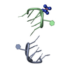
| |||||||||
|---|---|---|---|---|---|---|---|---|---|---|
| 1 | 
| |||||||||
| Unit cell |
| |||||||||
| Components on special symmetry positions |
|
-Components
| #1: DNA chain | Mass: 2113.410 Da / Num. of mol.: 2 / Source method: obtained synthetically #2: Chemical | ChemComp-NCO / |   Mass: 161.116 Da / Num. of mol.: 1 / Source method: obtained synthetically / Formula: CoH18N6 Mass: 161.116 Da / Num. of mol.: 1 / Source method: obtained synthetically / Formula: CoH18N6#3: Water | ChemComp-HOH / |  Mass: 18.015 Da / Num. of mol.: 38 / Source method: isolated from a natural source / Formula: H2O Mass: 18.015 Da / Num. of mol.: 38 / Source method: isolated from a natural source / Formula: H2O |
|---|
-Experimental details
-Experiment
| Experiment | Method: X-RAY DIFFRACTION / Number of used crystals: 1 |
|---|
- Sample preparation
| Crystal | Density Matthews: 1.77 Å3/Da / Density % sol: 30.67 % | ||||||||||||||||||||||||||||||||||||||||||||||||
|---|---|---|---|---|---|---|---|---|---|---|---|---|---|---|---|---|---|---|---|---|---|---|---|---|---|---|---|---|---|---|---|---|---|---|---|---|---|---|---|---|---|---|---|---|---|---|---|---|---|
| Crystal grow | Temperature: 293 K / Method: vapor diffusion, sitting drop / pH: 6.6 Details: Cobalt Hexammine, Cacodylate, MPD, NaCl, KCl, pH 6.6, VAPOR DIFFUSION, SITTING DROP, temperature 293K | ||||||||||||||||||||||||||||||||||||||||||||||||
| Components of the solutions |
| ||||||||||||||||||||||||||||||||||||||||||||||||
| Crystal grow | *PLUS Temperature: 290 K | ||||||||||||||||||||||||||||||||||||||||||||||||
| Components of the solutions | *PLUS
|
-Data collection
| Diffraction | Mean temperature: 100 K |
|---|---|
| Diffraction source | Source: SYNCHROTRON / Site: EMBL/DESY, HAMBURG  / Beamline: X11 / Wavelength: 0.811 / Wavelength: 0.811 Å / Beamline: X11 / Wavelength: 0.811 / Wavelength: 0.811 Å |
| Detector | Type: MARRESEARCH / Detector: CCD / Date: May 19, 2002 |
| Radiation | Protocol: SINGLE WAVELENGTH / Monochromatic (M) / Laue (L): M / Scattering type: x-ray |
| Radiation wavelength | Wavelength: 0.811 Å / Relative weight: 1 |
| Reflection | Resolution: 1.1→29.62 Å / Num. obs: 12514 / % possible obs: 99.3 % / Redundancy: 7.7 % / Biso Wilson estimate: 18.213 Å2 / Rmerge(I) obs: 0.11 / Net I/σ(I): 10.96 |
| Reflection shell | Resolution: 1.1→1.2 Å / Redundancy: 6.59 % / Rmerge(I) obs: 0.37 / Mean I/σ(I) obs: 5.24 / Num. unique all: 2803 / % possible all: 99.4 |
| Reflection | *PLUS Highest resolution: 1.1 Å / Lowest resolution: 20 Å / Num. obs: 2803 / Redundancy: 7.7 % / Num. measured all: 12514 |
| Reflection shell | *PLUS Highest resolution: 1.1 Å / Lowest resolution: 1.2 Å / % possible obs: 99.4 % / Redundancy: 6.64 % |
- Processing
| Software |
| |||||||||||||||||||||||||||||||||
|---|---|---|---|---|---|---|---|---|---|---|---|---|---|---|---|---|---|---|---|---|---|---|---|---|---|---|---|---|---|---|---|---|---|---|
| Refinement | Method to determine structure: MOLECULAR REPLACEMENT Starting model: UDG028 Resolution: 1.1→20 Å / Num. parameters: 2924 / Num. restraintsaints: 2847 / Cross valid method: FREE R / σ(F): 0 / Stereochemistry target values: ENGH AND HUBER / Details: ANISOTROPIC REFINEMENT REDUCED FREE R (NO CUTOFF)
| |||||||||||||||||||||||||||||||||
| Refine analyze | Num. disordered residues: 0 / Occupancy sum hydrogen: 175 / Occupancy sum non hydrogen: 322 | |||||||||||||||||||||||||||||||||
| Refinement step | Cycle: LAST / Resolution: 1.1→20 Å
| |||||||||||||||||||||||||||||||||
| Refine LS restraints |
| |||||||||||||||||||||||||||||||||
| Software | *PLUS Name: SHELXL / Version: 97 / Classification: refinement | |||||||||||||||||||||||||||||||||
| Refinement | *PLUS Lowest resolution: 20 Å / Rfactor Rfree: 0.224 | |||||||||||||||||||||||||||||||||
| Solvent computation | *PLUS | |||||||||||||||||||||||||||||||||
| Displacement parameters | *PLUS | |||||||||||||||||||||||||||||||||
| LS refinement shell | *PLUS Highest resolution: 1.1 Å / Lowest resolution: 1.2 Å / Rfactor Rfree: 0.2173 / Rfactor Rwork: 0.208 |
