 Movie
Movie Controller

[English] 日本語
 Yorodumi
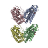
- PDB-1m7r: Crystal Structure of Myotubularin-related Protein-2 (MTMR2) Compl... -
+ Open data

- Basic information
| Entry | Database: PDB / ID: 1m7r | ||||||
|---|---|---|---|---|---|---|---|
| Title | Crystal Structure of Myotubularin-related Protein-2 (MTMR2) Complexed with Phosphate | ||||||
 Components Components | Myotubularin-related Protein-2 | ||||||
 Keywords Keywords | HYDROLASE / Protein-phosphate complex | ||||||
| Function / homology |  Function and homology information Function and homology informationnegative regulation of receptor catabolic process / phosphatidylinositol-3,5-bisphosphate 3-phosphatase / Synthesis of PIPs at the ER membrane / phosphatidylinositol-3,5-bisphosphate 3-phosphatase activity / regulation of phosphatidylinositol dephosphorylation / phosphatidylinositol-3-phosphate phosphatase activity / myelin assembly / positive regulation of early endosome to late endosome transport / Synthesis of PIPs at the late endosome membrane / Synthesis of PIPs at the early endosome membrane ...negative regulation of receptor catabolic process / phosphatidylinositol-3,5-bisphosphate 3-phosphatase / Synthesis of PIPs at the ER membrane / phosphatidylinositol-3,5-bisphosphate 3-phosphatase activity / regulation of phosphatidylinositol dephosphorylation / phosphatidylinositol-3-phosphate phosphatase activity / myelin assembly / positive regulation of early endosome to late endosome transport / Synthesis of PIPs at the late endosome membrane / Synthesis of PIPs at the early endosome membrane / negative regulation of myelination / phosphatidylinositol dephosphorylation / negative regulation of excitatory postsynaptic potential / negative regulation of endocytosis / phosphatidylinositol biosynthetic process / negative regulation of receptor internalization / dendritic spine maintenance / vacuolar membrane / Synthesis of PIPs at the plasma membrane / neuron development / synaptic membrane / synaptic vesicle / early endosome membrane / dendritic spine / postsynaptic density / axon / dendrite / perinuclear region of cytoplasm / extracellular exosome / membrane / identical protein binding / nucleus / cytoplasm / cytosol Similarity search - Function | ||||||
| Biological species |  Homo sapiens (human) Homo sapiens (human) | ||||||
| Method |  X-RAY DIFFRACTION / SYNCHROTRON / MAD / Resolution: 2.6 Å X-RAY DIFFRACTION / SYNCHROTRON / MAD / Resolution: 2.6 Å | ||||||
 Authors Authors | Begley, M.J. / Taylor, G.S. / Kim, S.-A. / Veine, D.M. / Dixon, J.E. / Stuckey, J.A. | ||||||
 Citation Citation | Journal: Mol.Cell / Year: 2003 Title: Crystal structure of a phosphoinositide phosphatase, MTMR2: insights into myotubular myopathy and Charcot-Marie-Tooth syndrome Authors: Begley, M.J. / Taylor, G.S. / Kim, S.-A. / Veine, D.M. / Dixon, J.E. / Stuckey, J.A. | ||||||
| History |
|
- Structure visualization
| Structure viewer | Molecule: MolmilJmol/JSmol |
|---|
- Downloads & links
-Download
| PDBx/mmCIF format | 1m7r.cif.gz | 224.5 KB | Display | PDBx/mmCIF format |
|---|---|---|---|---|
| PDB format | pdb1m7r.ent.gz | 177.9 KB | Display | PDB format |
| PDBx/mmJSON format | 1m7r.json.gz | Tree view | PDBx/mmJSON format | |
| Others |  Other downloads Other downloads |
-Validation report
| Arichive directory | https://data.pdbj.org/pub/pdb/validation_reports/m7/1m7rftp://data.pdbj.org/pub/pdb/validation_reports/m7/1m7r | HTTPS FTP |
|---|
-Related structure data


-Links
 PDBj
PDBj


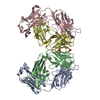
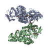
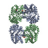
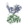
- Assembly
| Deposited unit | 
| ||||||||
|---|---|---|---|---|---|---|---|---|---|
| 1 |
| ||||||||
| Unit cell |
|
-Components
| #1: Protein | Mass: 75006.742 Da / Num. of mol.: 2 / Fragment: PH and Phosphatase Domains (Residues 1-643) / Mutation: C417S Source method: isolated from a genetically manipulated source Source: (gene. exp.) Homo sapiens (human) / Gene: MTMR2 / Plasmid: PET / Species (production host): Escherichia coli / Production host:  References: UniProt: Q13614, phosphatidylinositol-3-phosphatase #2: Chemical | ChemComp-PO4 /   Mass: 94.971 Da / Num. of mol.: 4 / Source method: obtained synthetically / Formula: PO4 Mass: 94.971 Da / Num. of mol.: 4 / Source method: obtained synthetically / Formula: PO4#3: Water | ChemComp-HOH / |  Mass: 18.015 Da / Num. of mol.: 234 / Source method: isolated from a natural source / Formula: H2O Mass: 18.015 Da / Num. of mol.: 234 / Source method: isolated from a natural source / Formula: H2O |
|---|
-Experimental details
-Experiment
| Experiment | Method: X-RAY DIFFRACTION / Number of used crystals: 1 |
|---|
- Sample preparation
| Crystal | Density Matthews: 0.968 Å3/Da / Density % sol: 31.93 % | |||||||||||||||||||||||||||||||||||||||||||||||||||||||||||||||
|---|---|---|---|---|---|---|---|---|---|---|---|---|---|---|---|---|---|---|---|---|---|---|---|---|---|---|---|---|---|---|---|---|---|---|---|---|---|---|---|---|---|---|---|---|---|---|---|---|---|---|---|---|---|---|---|---|---|---|---|---|---|---|---|---|
| Crystal grow | Method: vapor diffusion, hanging drop / pH: 7.5 Details: PEG 35000, Hepes, pH 7.5, VAPOR DIFFUSION, HANGING DROP, temperature 100.0K | |||||||||||||||||||||||||||||||||||||||||||||||||||||||||||||||
| Crystal grow | *PLUS Temperature: 20 ℃ / pH: 8 / Method: vapor diffusion, hanging drop | |||||||||||||||||||||||||||||||||||||||||||||||||||||||||||||||
| Components of the solutions | *PLUS
|
-Data collection
| Diffraction | Mean temperature: 100 K |
|---|---|
| Diffraction source | Source: SYNCHROTRON / Site: APS  / Beamline: 5ID-B / Wavelength: 0.98 Å / Beamline: 5ID-B / Wavelength: 0.98 Å |
| Detector | Type: MARRESEARCH / Detector: CCD / Date: Aug 20, 2001 / Details: Mirrors |
| Radiation | Monochromator: SI 111 Channel / Protocol: MAD / Monochromatic (M) / Laue (L): M / Scattering type: x-ray |
| Radiation wavelength | Wavelength: 0.98 Å / Relative weight: 1 |
| Reflection | Resolution: 2.6→30 Å / Num. all: 39438 / Num. obs: 39438 / % possible obs: 100 % / Observed criterion σ(F): 0 / Observed criterion σ(I): 0 / Redundancy: 8 % / Biso Wilson estimate: 40.2 Å2 / Rsym value: 0.07 / Net I/σ(I): 11.9 |
| Reflection shell | Resolution: 2.6→2.76 Å / Mean I/σ(I) obs: 5.5 / Rsym value: 0.282 / % possible all: 95.6 |
| Reflection | *PLUS Num. measured all: 459390 / Rmerge(I) obs: 0.08 |
| Reflection shell | *PLUS % possible obs: 100 % / Rmerge(I) obs: 0.254 |
- Processing
| Software |
| ||||||||||||||||||||||||||||||||||||
|---|---|---|---|---|---|---|---|---|---|---|---|---|---|---|---|---|---|---|---|---|---|---|---|---|---|---|---|---|---|---|---|---|---|---|---|---|---|
| Refinement | Method to determine structure: MAD / Resolution: 2.6→8 Å / Rfactor Rfree error: 0.005 / Isotropic thermal model: Restrained / Cross valid method: THROUGHOUT / σ(F): 0 / σ(I): 0 / Stereochemistry target values: Engh & Huber
| ||||||||||||||||||||||||||||||||||||
| Solvent computation | Solvent model: FLAT MODEL / Bsol: 57.2692 Å2 / ksol: 0.455054 e/Å3 | ||||||||||||||||||||||||||||||||||||
| Displacement parameters | Biso mean: 41.51 Å2
| ||||||||||||||||||||||||||||||||||||
| Refine analyze |
| ||||||||||||||||||||||||||||||||||||
| Refinement step | Cycle: LAST / Resolution: 2.6→8 Å
| ||||||||||||||||||||||||||||||||||||
| Refine LS restraints |
| ||||||||||||||||||||||||||||||||||||
| LS refinement shell | Resolution: 2.6→2.76 Å / Rfactor Rfree error: 0.017 / Total num. of bins used: 6
| ||||||||||||||||||||||||||||||||||||
| Xplor file | Serial no: 1 / Param file: protein_rep.param / Topol file: protein.top | ||||||||||||||||||||||||||||||||||||
| Refinement | *PLUS Highest resolution: 2.6 Å / Lowest resolution: 8 Å / Rfactor Rfree: 0.287 / Rfactor Rwork: 0.212 | ||||||||||||||||||||||||||||||||||||
| Solvent computation | *PLUS | ||||||||||||||||||||||||||||||||||||
| Displacement parameters | *PLUS | ||||||||||||||||||||||||||||||||||||
| Refine LS restraints | *PLUS
|
