 Movie
Movie Controller

[English] 日本語
 Yorodumi
- PDB-1m2f: Solution structure of the N-terminal domain of Synechococcus elon... -
+ Open data

- Basic information
| Entry | Database: PDB / ID: 1m2f | ||||||
|---|---|---|---|---|---|---|---|
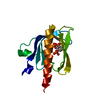
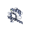
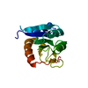
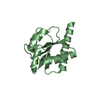
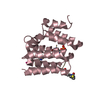
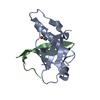
| Title | Solution structure of the N-terminal domain of Synechococcus elongatus KaiA (KaiA135N); Family of 25 structures | ||||||
 Components Components | KaiA | ||||||
 Keywords Keywords | CIRCADIAN CLOCK PROTEIN / ALPHA-BETA-ALPHA SANDWICH | ||||||
| Function / homology |  Function and homology information Function and homology informationdetection of redox state / entrainment of circadian clock / positive regulation of circadian rhythm / circadian rhythm / identical protein binding Similarity search - Function | ||||||
| Biological species |  Synechococcus elongatus (bacteria) Synechococcus elongatus (bacteria) | ||||||
| Method | SOLUTION NMR / Distance geometry, Simulated annealing regularization, Simulated annealing refinement | ||||||
 Authors Authors | Williams, S.B. / Vakonakis, I. / Golden, S.S. / LiWang, A.C. | ||||||
 Citation Citation | Journal: Proc.Natl.Acad.Sci.USA / Year: 2002 Title: Structure and Function from the Circadian Clock Protein KaiA of Synechococcus elongatus: A potential Clock Input Mechanism Authors: Williams, S.B. / Vakonakis, I. / Golden, S.S. / LiWang, A.C. | ||||||
| History |
|
- Structure visualization
| Structure viewer | Molecule: MolmilJmol/JSmol |
|---|
- Downloads & links
-Download
| PDBx/mmCIF format | 1m2f.cif.gz | 1009 KB | Display | PDBx/mmCIF format |
|---|---|---|---|---|
| PDB format | pdb1m2f.ent.gz | 845.9 KB | Display | PDB format |
| PDBx/mmJSON format | 1m2f.json.gz | Tree view | PDBx/mmJSON format | |
| Others |  Other downloads Other downloads |
-Validation report
| Arichive directory | https://data.pdbj.org/pub/pdb/validation_reports/m2/1m2fftp://data.pdbj.org/pub/pdb/validation_reports/m2/1m2f | HTTPS FTP |
|---|
-Related structure data
| Related structure data |  1m2eC C: citing same article ( |
|---|---|
| Similar structure data | |
| Other databases |
|


-Links
 PDBj
PDBj

- Assembly
| Deposited unit | 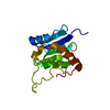
| |||||||||
|---|---|---|---|---|---|---|---|---|---|---|
| 1 |
| |||||||||
| NMR ensembles |
|
-Components
| #1: Protein | Mass: 15077.117 Da / Num. of mol.: 1 / Fragment: N-terminal domain (Residues 1-135) Source method: isolated from a genetically manipulated source Source: (gene. exp.) Synechococcus elongatus (bacteria) / Gene: KaiA / Plasmid: pET32a / Species (production host): Escherichia coli / Production host: |
|---|
-Experimental details
-Experiment
| Experiment | Method: SOLUTION NMR | ||||||||||||||||||||||||||||
|---|---|---|---|---|---|---|---|---|---|---|---|---|---|---|---|---|---|---|---|---|---|---|---|---|---|---|---|---|---|
| NMR experiment |
| ||||||||||||||||||||||||||||
| NMR details | Text: Submission corresponds to family of 25-low-energy-structures. The 1H, 15N and 13C chemical shifts of KaiA135N are deposited at the BMRB database (http://www.bmrb.wisc.edu) under the accession number 5031. |
- Sample preparation
| Details |
| ||||||||||||
|---|---|---|---|---|---|---|---|---|---|---|---|---|---|
| Sample conditions | Ionic strength: 0.17 / pH: 7 / Pressure: ambient / Temperature: 298 K | ||||||||||||
| Crystal grow | *PLUS Method: other / Details: NMR |
-NMR measurement
| Radiation | Protocol: SINGLE WAVELENGTH / Monochromatic (M) / Laue (L): M |
|---|---|
| Radiation wavelength | Relative weight: 1 |
| NMR spectrometer | Type: Varian INOVA / Manufacturer: Varian / Model: INOVA / Field strength: 600 MHz |
- Processing
| NMR software |
| ||||||||||||||||||||
|---|---|---|---|---|---|---|---|---|---|---|---|---|---|---|---|---|---|---|---|---|---|
| Refinement | Method: Distance geometry, Simulated annealing regularization, Simulated annealing refinement Software ordinal: 1 Details: The structures are based on 2034 restraints: 1816 are NOE-derived distance constraints, 187 dihedral angle restraints, 31 distance restraints from hydrogen bonds | ||||||||||||||||||||
| NMR representative | Selection criteria: closest to the average | ||||||||||||||||||||
| NMR ensemble | Conformer selection criteria: structures with the lowest energy Conformers calculated total number: 50 / Conformers submitted total number: 25 |
 NMRPipe