 Movie
Movie Controller

[English] 日本語
 Yorodumi
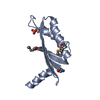
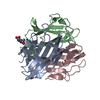
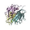
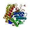
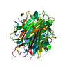
- PDB-1lxj: X-RAY STRUCTURE OF YBL001c NORTHEAST STRUCTURAL GENOMICS (NESG) C... -
+ Open data

- Basic information
| Entry | Database: PDB / ID: 1lxj | ||||||
|---|---|---|---|---|---|---|---|
| Title | X-RAY STRUCTURE OF YBL001c NORTHEAST STRUCTURAL GENOMICS (NESG) CONSORTIUM TARGET YTYst72 | ||||||
 Components Components | HYPOTHETICAL 11.5KDA PROTEIN IN HTB2-NTH2 INTERGENIC REGION | ||||||
 Keywords Keywords | STRUCTURAL GENOMICS / UNKNOWN FUNCTION / Hypothetical protein / HTB2-NTH2 intergenic region / PSI / Protein Structure Initiative / Northeast Structural Genomics Consortium / NESG | ||||||
| Function / homology |  Function and homology information Function and homology information | ||||||
| Biological species |  | ||||||
| Method |  X-RAY DIFFRACTION / SYNCHROTRON / SAD / Resolution: 1.8 Å X-RAY DIFFRACTION / SYNCHROTRON / SAD / Resolution: 1.8 Å | ||||||
 Authors Authors | Tao, X. / Khayat, R. / Christendat, D. / Savchenko, A. / Xu, X. / Edwards, A. / Arrowsmith, C.H. / Tong, L. / Northeast Structural Genomics Consortium (NESG) | ||||||
 Citation Citation | Journal: Proteins / Year: 2003 Title: CRYSTAL STRUCTURES OF MTH1187 AND ITS YEAST ORTHOLOG YBL001C Authors: Tao, X. / Khayat, R. / Christendat, D. / Savchenko, A. / Xu, X. / Goldsmith-Fischman, S. / Honig, B. / Edwards, A. / Arrowsmith, C.H. / Tong, L. | ||||||
| History |
|
- Structure visualization
| Structure viewer | Molecule: MolmilJmol/JSmol |
|---|
- Downloads & links
-Download
| PDBx/mmCIF format | 1lxj.cif.gz | 38.6 KB | Display | PDBx/mmCIF format |
|---|---|---|---|---|
| PDB format | pdb1lxj.ent.gz | 26.1 KB | Display | PDB format |
| PDBx/mmJSON format | 1lxj.json.gz | Tree view | PDBx/mmJSON format | |
| Others |  Other downloads Other downloads |
-Validation report
| Arichive directory | https://data.pdbj.org/pub/pdb/validation_reports/lx/1lxjftp://data.pdbj.org/pub/pdb/validation_reports/lx/1lxj | HTTPS FTP |
|---|
-Related structure data


-Links
 PDBj
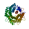
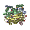
PDBj- Assembly
| Deposited unit | 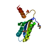
| ||||||||
|---|---|---|---|---|---|---|---|---|---|
| 1 | 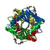
| ||||||||
| Unit cell |
|
-Components
| #1: Protein | Mass: 11676.963 Da / Num. of mol.: 1 Source method: isolated from a genetically manipulated source Source: (gene. exp.) Production host:  |
|---|---|
| #2: Chemical | ChemComp-SO4 /   Mass: 96.063 Da / Num. of mol.: 1 / Source method: obtained synthetically / Formula: SO4 Mass: 96.063 Da / Num. of mol.: 1 / Source method: obtained synthetically / Formula: SO4 |
| #3: Water | ChemComp-HOH /  Mass: 18.015 Da / Num. of mol.: 223 / Source method: isolated from a natural source / Formula: H2O Mass: 18.015 Da / Num. of mol.: 223 / Source method: isolated from a natural source / Formula: H2O |
| Has protein modification | Y |
-Experimental details
-Experiment
| Experiment | Method: X-RAY DIFFRACTION / Number of used crystals: 1 |
|---|
- Sample preparation
| Crystal | Density Matthews: 3.25 Å3/Da / Density % sol: 62.1 % | |||||||||||||||||||||||||||||||||||||||||||||||||
|---|---|---|---|---|---|---|---|---|---|---|---|---|---|---|---|---|---|---|---|---|---|---|---|---|---|---|---|---|---|---|---|---|---|---|---|---|---|---|---|---|---|---|---|---|---|---|---|---|---|---|
| Crystal grow | Temperature: 293 K / Method: vapor diffusion, hanging drop / pH: 7 Details: Ammonium Sulfate, 2-METHYL-2,4-PENTANEDIOL, pH 7.0, VAPOR DIFFUSION, HANGING DROP, temperature 293K | |||||||||||||||||||||||||||||||||||||||||||||||||
| Crystal grow | *PLUS Temperature: 4 ℃ / pH: 8.5 / Method: vapor diffusion | |||||||||||||||||||||||||||||||||||||||||||||||||
| Components of the solutions | *PLUS
|
-Data collection
| Diffraction | Mean temperature: 100 K |
|---|---|
| Diffraction source | Source: SYNCHROTRON / Site: NSLS  / Beamline: X4A / Wavelength: 0.97895 Å / Beamline: X4A / Wavelength: 0.97895 Å |
| Detector | Type: ADSC QUANTUM 4 / Detector: CCD / Date: May 4, 2002 |
| Radiation | Monochromator: Si 111 CHANNEL / Protocol: SINGLE WAVELENGTH / Monochromatic (M) / Laue (L): M / Scattering type: x-ray |
| Radiation wavelength | Wavelength: 0.97895 Å / Relative weight: 1 |
| Reflection | Resolution: 1.8→30 Å / Num. obs: 12726 / % possible obs: 86.8 % / Observed criterion σ(F): 0 / Observed criterion σ(I): 0 / Biso Wilson estimate: 16.5 Å2 |
| Reflection shell | Resolution: 1.8→1.86 Å / % possible all: 85.7 |
| Reflection | *PLUS Highest resolution: 1.8 Å / Num. measured all: 141589 / Rmerge(I) obs: 0.096 |
- Processing
| Software |
| ||||||||||||||||||||||||||||||||||||
|---|---|---|---|---|---|---|---|---|---|---|---|---|---|---|---|---|---|---|---|---|---|---|---|---|---|---|---|---|---|---|---|---|---|---|---|---|---|
| Refinement | Method to determine structure: SAD / Resolution: 1.8→19.99 Å / Rfactor Rfree error: 0.008 / Data cutoff high absF: 4329413.23 / Data cutoff high rms absF: 4329413.23 / Data cutoff low absF: 0 / Isotropic thermal model: RESTRAINED / Cross valid method: THROUGHOUT / σ(F): 2 / Stereochemistry target values: Engh & Huber
| ||||||||||||||||||||||||||||||||||||
| Solvent computation | Bsol: 52.0701 Å2 / ksol: 0.351892 e/Å3 | ||||||||||||||||||||||||||||||||||||
| Displacement parameters | Biso mean: 30.8 Å2
| ||||||||||||||||||||||||||||||||||||
| Refine analyze |
| ||||||||||||||||||||||||||||||||||||
| Refinement step | Cycle: LAST / Resolution: 1.8→19.99 Å
| ||||||||||||||||||||||||||||||||||||
| Refine LS restraints |
| ||||||||||||||||||||||||||||||||||||
| LS refinement shell | Resolution: 1.8→1.91 Å / Rfactor Rfree error: 0.028 / Total num. of bins used: 6
| ||||||||||||||||||||||||||||||||||||
| Xplor file |
| ||||||||||||||||||||||||||||||||||||
| Refinement | *PLUS Highest resolution: 1.8 Å / Lowest resolution: 20 Å / Num. reflection obs: 13776 | ||||||||||||||||||||||||||||||||||||
| Solvent computation | *PLUS | ||||||||||||||||||||||||||||||||||||
| Displacement parameters | *PLUS | ||||||||||||||||||||||||||||||||||||
| Refine LS restraints | *PLUS
|
