 Movie
Movie Controller


+ Open data

- Basic information
| Entry | Database: PDB / ID: 1lt3 | |||||||||
|---|---|---|---|---|---|---|---|---|---|---|
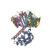
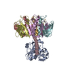
| Title | HEAT-LABILE ENTEROTOXIN DOUBLE MUTANT N40C/G166C | |||||||||
 Components Components | (HEAT-LABILE ENTEROTOXIN) x 2 | |||||||||
 Keywords Keywords | ENTEROTOXIN | |||||||||
| Function / homology |  Function and homology information Function and homology informationSecretion of toxins / toxin activity / killing of cells of another organism / : / extracellular region Similarity search - Function | |||||||||
| Biological species |  | |||||||||
| Method |  X-RAY DIFFRACTION / ISOMORPHOUS MOLECULAR REPLACEMENT / Resolution: 2 Å X-RAY DIFFRACTION / ISOMORPHOUS MOLECULAR REPLACEMENT / Resolution: 2 Å | |||||||||
 Authors Authors | Van Den Akker, F. / Hol, W.G.J. | |||||||||
 Citation Citation | Journal: Protein Sci. / Year: 1997 Title: Crystal structure of heat-labile enterotoxin from Escherichia coli with increased thermostability introduced by an engineered disulfide bond in the A subunit. Authors: van den Akker, F. / Feil, I.K. / Roach, C. / Platas, A.A. / Merritt, E.A. / Hol, W.G. #1: Journal: J.Mol.Biol. / Year: 1993Title: Refined Structure of Escherichia Coli Heat-Labile Enterotoxin, a Close Relative of Cholera Toxin Authors: Sixma, T.K. / Kalk, K.H. / Van Zanten, B.A. / Dauter, Z. / Kingma, J. / Witholt, B. / Hol, W.G. #2: Journal: Nature / Year: 1991Title: Crystal Structure of a Cholera Toxin-Related Heat-Labile Enterotoxin from E. Coli Authors: Sixma, T.K. / Pronk, S.E. / Kalk, K.H. / Wartna, E.S. / Van Zanten, B.A. / Witholt, B. / Hol, W.G. | |||||||||
| History |
|
- Structure visualization
| Structure viewer | Molecule: MolmilJmol/JSmol |
|---|
- Downloads & links
-Download
| PDBx/mmCIF format | 1lt3.cif.gz | 159.1 KB | Display | PDBx/mmCIF format |
|---|---|---|---|---|
| PDB format | pdb1lt3.ent.gz | 126.7 KB | Display | PDB format |
| PDBx/mmJSON format | 1lt3.json.gz | Tree view | PDBx/mmJSON format | |
| Others |  Other downloads Other downloads |
-Validation report
| Arichive directory | https://data.pdbj.org/pub/pdb/validation_reports/lt/1lt3ftp://data.pdbj.org/pub/pdb/validation_reports/lt/1lt3 | HTTPS FTP |
|---|
-Related structure data
| Related structure data | 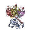 1lttS S: Starting model for refinement |
|---|---|
| Similar structure data |


-Links
 PDBj
PDBj


- Assembly
| Deposited unit | 
| ||||||||
|---|---|---|---|---|---|---|---|---|---|
| 1 |
| ||||||||
| Unit cell |
| ||||||||
| Details | ASYMMETRIC UNIT CONTAINS ONE AB5 TOXIN HEXAMER. THE A SUBUNIT CONTAINS TWO FRAGMENTS LINKED BY A DISORDERED LOOP. THESE 2 FRAGMENTS ARE CONVENTIONALLY REFERRED TO AS A1 AND A2. FRAGMENTS A1 AND A2 ARE COVALENTLY LINKED IN THE LATENT TOXIN AND ARE PROTEOLYTICALLY CLEAVED UPON ACTIVATION. |
-Components
| #1: Protein | Mass: 11807.539 Da / Num. of mol.: 5 / Fragment: HOLOTOXIN / Mutation: N40C, G166C Source method: isolated from a genetically manipulated source Details: LACTOSE BOUND / Source: (gene. exp.) #2: Protein | | Mass: 27886.736 Da / Num. of mol.: 1 / Fragment: HOLOTOXIN / Mutation: N40C, G166C Source method: isolated from a genetically manipulated source Details: LACTOSE BOUND / Source: (gene. exp.) #3: Polysaccharide | beta-D-galactopyranose-(1-4)-beta-D-glucopyranose / beta-lactose   Source method: isolated from a genetically manipulated source Details: oligosaccharide / References: beta-lactose Has protein modification | Y | |
|---|
-Experimental details
-Experiment
| Experiment | Method: X-RAY DIFFRACTION / Number of used crystals: 1 |
|---|
- Sample preparation
| Crystal | Density Matthews: 2.23 Å3/Da / Density % sol: 44.94 % Description: NO MOLECULAR REPLACEMENT SEARCH NEEDED TO BE PERFORMED SINCE THE SPACE GROUP AND CELL DIMENSIONS WERE IDENTICAL TO THE STARTING 1LTT STRUCTURE. | ||||||||||||||||||||||||||||||||||||||||||||||||||||||||
|---|---|---|---|---|---|---|---|---|---|---|---|---|---|---|---|---|---|---|---|---|---|---|---|---|---|---|---|---|---|---|---|---|---|---|---|---|---|---|---|---|---|---|---|---|---|---|---|---|---|---|---|---|---|---|---|---|---|
| Crystal grow | Method: 3 layer capillary method / pH: 7.5 Details: PROTEIN WAS CRYSTALLIZED FROM 5% PEG 6000, 100 MM NACL, 1 MM EDTA, 75 MM LACTOSE, 100 MM TRIS PH 7.5 USING THE 3 LAYER CAPPILARY METHOD, 3 layer capillary method | ||||||||||||||||||||||||||||||||||||||||||||||||||||||||
| Crystal grow | *PLUS Method: three-layer capillary method | ||||||||||||||||||||||||||||||||||||||||||||||||||||||||
| Components of the solutions | *PLUS
|
-Data collection
| Diffraction | Mean temperature: 295 K |
|---|---|
| Diffraction source | Source: ROTATING ANODE / Type: RIGAKU RUH2R / Wavelength: 1.5418 |
| Detector | Type: RIGAKU RAXIS IV / Detector: IMAGE PLATE / Date: Sep 1, 1996 / Details: MIRRORS |
| Radiation | Monochromator: NI FILTER / Monochromatic (M) / Laue (L): M / Scattering type: x-ray |
| Radiation wavelength | Wavelength: 1.5418 Å / Relative weight: 1 |
| Reflection | Resolution: 2→100 Å / Num. obs: 48385 / % possible obs: 90.5 % / Observed criterion σ(I): 1 / Redundancy: 2.9 % / Rsym value: 0.053 / Net I/σ(I): 17 |
| Reflection shell | Resolution: 2→2.07 Å / Mean I/σ(I) obs: 4.5 / Rsym value: 0.175 / % possible all: 72 |
| Reflection | *PLUS Rmerge(I) obs: 0.053 |
| Reflection shell | *PLUS % possible obs: 72.3 % / Rmerge(I) obs: 0.175 |
- Processing
| Software |
| ||||||||||||||||||||||||||||||||||||||||||||||||||||||||||||
|---|---|---|---|---|---|---|---|---|---|---|---|---|---|---|---|---|---|---|---|---|---|---|---|---|---|---|---|---|---|---|---|---|---|---|---|---|---|---|---|---|---|---|---|---|---|---|---|---|---|---|---|---|---|---|---|---|---|---|---|---|---|
| Refinement | Method to determine structure: ISOMORPHOUS MOLECULAR REPLACEMENT Starting model: PDB ENTRY 1LTT Resolution: 2→10 Å / Cross valid method: THROUGHOUT / σ(F): 2 Details: RESIDUES 1 - 3, 189 - 195 AND 237 - 240 OF THE A SUBUNIT ARE OMITTED FROM THE STRUCTURE BECAUSE OF POOR ELECTRON DENSITY GLY A 188 IS THE LAST RESIDUE BEFORE GAP (RESIDUES A 189 - A 195 ARE ...Details: RESIDUES 1 - 3, 189 - 195 AND 237 - 240 OF THE A SUBUNIT ARE OMITTED FROM THE STRUCTURE BECAUSE OF POOR ELECTRON DENSITY GLY A 188 IS THE LAST RESIDUE BEFORE GAP (RESIDUES A 189 - A 195 ARE DISORDERED). LEU A 240 IS THE LAST RESIDUE BUT RESIDUES A 237 - A 240 ARE DISORDERED.
| ||||||||||||||||||||||||||||||||||||||||||||||||||||||||||||
| Displacement parameters | Biso mean: 25.9 Å2 | ||||||||||||||||||||||||||||||||||||||||||||||||||||||||||||
| Refinement step | Cycle: LAST / Resolution: 2→10 Å
| ||||||||||||||||||||||||||||||||||||||||||||||||||||||||||||
| Refine LS restraints |
| ||||||||||||||||||||||||||||||||||||||||||||||||||||||||||||
| LS refinement shell | Resolution: 2→2.07 Å / Total num. of bins used: 10
| ||||||||||||||||||||||||||||||||||||||||||||||||||||||||||||
| Xplor file |
| ||||||||||||||||||||||||||||||||||||||||||||||||||||||||||||
| Software | *PLUS Name: X-PLOR / Version: 3.1 / Classification: refinement | ||||||||||||||||||||||||||||||||||||||||||||||||||||||||||||
| Refinement | *PLUS Num. reflection obs: 45930 | ||||||||||||||||||||||||||||||||||||||||||||||||||||||||||||
| Solvent computation | *PLUS | ||||||||||||||||||||||||||||||||||||||||||||||||||||||||||||
| Displacement parameters | *PLUS | ||||||||||||||||||||||||||||||||||||||||||||||||||||||||||||
| Refine LS restraints | *PLUS
|
