 Movie
Movie Controller

[English] 日本語
 Yorodumi
- PDB-1lqs: CRYSTAL STRUCTURE OF HUMAN CYTOMEGALOVIRUS IL-10 BOUND TO SOLUBLE... -
+ Open data

- Basic information
| Entry | Database: PDB / ID: 1lqs | ||||||
|---|---|---|---|---|---|---|---|
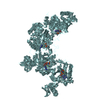
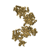
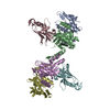
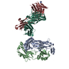
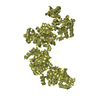
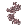
| Title | CRYSTAL STRUCTURE OF HUMAN CYTOMEGALOVIRUS IL-10 BOUND TO SOLUBLE HUMAN IL-10R1 | ||||||
 Components Components |
| ||||||
 Keywords Keywords | IMMUNE SYSTEM / interleukin 10 / helix bundle / receptor complex / molecular recognition / structure mimic | ||||||
| Function / homology |  Function and homology information Function and homology informationsymbiont-mediated suppression of host dendritic cell mediated immune response / interleukin-10 binding / interleukin-10 receptor activity / interleukin-10-mediated signaling pathway / intestinal epithelial structure maintenance / ubiquitin-dependent endocytosis / regulation of synapse organization / Interleukin-10 signaling / Nuclear events stimulated by ALK signaling in cancer / negative regulation of autophagy ...symbiont-mediated suppression of host dendritic cell mediated immune response / interleukin-10 binding / interleukin-10 receptor activity / interleukin-10-mediated signaling pathway / intestinal epithelial structure maintenance / ubiquitin-dependent endocytosis / regulation of synapse organization / Interleukin-10 signaling / Nuclear events stimulated by ALK signaling in cancer / negative regulation of autophagy / cytokine activity / positive regulation of receptor signaling pathway via JAK-STAT / negative regulation of inflammatory response / cytokine-mediated signaling pathway / signaling receptor activity / response to lipopolysaccharide / apical plasma membrane / cilium / symbiont-mediated suppression of host innate immune response / immune response / symbiont-mediated suppression of host type I interferon-mediated signaling pathway / extracellular space / nucleoplasm / plasma membrane / cytosol Similarity search - Function | ||||||
| Biological species |  Homo sapiens (human) Homo sapiens (human)  Human herpesvirus 5 Human herpesvirus 5 | ||||||
| Method |  X-RAY DIFFRACTION / MOLECULAR REPLACEMENT / Resolution: 2.7 Å X-RAY DIFFRACTION / MOLECULAR REPLACEMENT / Resolution: 2.7 Å | ||||||
 Authors Authors | Jones, B.C. / Logsdon, N.J. / Josephson, K. / Cook, J. / Barry, P.A. / Walter, M.R. | ||||||
 Citation Citation | Journal: Proc.Natl.Acad.Sci.USA / Year: 2002 Title: Crystal structure of human cytomegalovirus IL-10 bound to soluble human IL-10R1. Authors: Jones, B.C. / Logsdon, N.J. / Josephson, K. / Cook, J. / Barry, P.A. / Walter, M.R. | ||||||
| History |
|
- Structure visualization
| Structure viewer | Molecule: MolmilJmol/JSmol |
|---|
- Downloads & links
-Download
| PDBx/mmCIF format | 1lqs.cif.gz | 151.2 KB | Display | PDBx/mmCIF format |
|---|---|---|---|---|
| PDB format | pdb1lqs.ent.gz | 119.4 KB | Display | PDB format |
| PDBx/mmJSON format | 1lqs.json.gz | Tree view | PDBx/mmJSON format | |
| Others |  Other downloads Other downloads |
-Validation report
| Arichive directory | https://data.pdbj.org/pub/pdb/validation_reports/lq/1lqsftp://data.pdbj.org/pub/pdb/validation_reports/lq/1lqs | HTTPS FTP |
|---|
-Related structure data
| Related structure data | 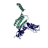 1j7vS S: Starting model for refinement |
|---|---|
| Similar structure data |


-Links
 PDBj
PDBj


- Assembly
| Deposited unit | 
| ||||||||
|---|---|---|---|---|---|---|---|---|---|
| 1 |
| ||||||||
| Unit cell |
|
-Components
| #1: Protein | Mass: 24506.551 Da / Num. of mol.: 2 / Fragment: EXTRACELLULAR DOMAIN, RESIDUES 22-235 / Mutation: N29Q,N53Q,N89Q,N133Q,N156Q,N168Q Source method: isolated from a genetically manipulated source Source: (gene. exp.) Homo sapiens (human) / Plasmid: pmtv5his / Production host:  #2: Protein | Mass: 18141.646 Da / Num. of mol.: 2 / Fragment: RESIDUES 20-176 Source method: isolated from a genetically manipulated source Source: (gene. exp.) Human herpesvirus 5 / Genus: Cytomegalovirus / Plasmid: pmtv5his / Production host: #3: Sugar |   Type: D-saccharide, beta linking / Mass: 221.208 Da / Num. of mol.: 2 Type: D-saccharide, beta linking / Mass: 221.208 Da / Num. of mol.: 2Source method: isolated from a genetically manipulated source Formula: C8H15NO6 #4: Water | ChemComp-HOH / |  Mass: 18.015 Da / Num. of mol.: 48 / Source method: isolated from a natural source / Formula: H2O Mass: 18.015 Da / Num. of mol.: 48 / Source method: isolated from a natural source / Formula: H2OHas protein modification | Y | |
|---|
-Experimental details
-Experiment
| Experiment | Method: X-RAY DIFFRACTION / Number of used crystals: 1 |
|---|
- Sample preparation
| Crystal | Density Matthews: 3.09 Å3/Da / Density % sol: 60.17 % | |||||||||||||||||||||||||||||||||||||||||||||||||
|---|---|---|---|---|---|---|---|---|---|---|---|---|---|---|---|---|---|---|---|---|---|---|---|---|---|---|---|---|---|---|---|---|---|---|---|---|---|---|---|---|---|---|---|---|---|---|---|---|---|---|
| Crystal grow | Temperature: 298 K / Method: vapor diffusion, hanging drop / pH: 6 Details: PEG-6000, 0.1M MgCl2, 100mM ADA, 0.75% PEG-400, VAPOR DIFFUSION, HANGING DROP, temperature 298K | |||||||||||||||||||||||||||||||||||||||||||||||||
| Crystal grow | *PLUS Temperature: 25 ℃ / pH: 8 | |||||||||||||||||||||||||||||||||||||||||||||||||
| Components of the solutions | *PLUS
|
-Data collection
| Diffraction | Mean temperature: 100 K |
|---|---|
| Diffraction source | Source: ROTATING ANODE / Type: RIGAKU RU200 / Wavelength: 1.54 Å |
| Detector | Type: RIGAKU RAXIS IV / Detector: IMAGE PLATE / Date: Oct 30, 2001 / Details: osmic mirrors |
| Radiation | Monochromator: osmic mirrors / Protocol: SINGLE WAVELENGTH / Monochromatic (M) / Laue (L): M / Scattering type: x-ray |
| Radiation wavelength | Wavelength: 1.54 Å / Relative weight: 1 |
| Reflection | Resolution: 2.7→50 Å / Num. all: 26564 / Num. obs: 26049 / Observed criterion σ(F): 0 / Observed criterion σ(I): -3 |
| Reflection shell | Resolution: 2.7→2.8 Å / Redundancy: 2.4 % / Rmerge(I) obs: 0.195 / % possible all: 0.74 |
| Reflection | *PLUS Lowest resolution: 50 Å / Num. obs: 26564 / % possible obs: 92.5 % / Redundancy: 2.8 % / Num. measured all: 65855 / Rmerge(I) obs: 0.056 |
| Reflection shell | *PLUS % possible obs: 73.7 % |
- Processing
| Software |
| ||||||||||||||||||||
|---|---|---|---|---|---|---|---|---|---|---|---|---|---|---|---|---|---|---|---|---|---|
| Refinement | Method to determine structure: MOLECULAR REPLACEMENT Starting model: PDB ENTRY 1J7V Resolution: 2.7→50 Å / σ(F): 0 / Stereochemistry target values: Engh & Huber
| ||||||||||||||||||||
| Refinement step | Cycle: LAST / Resolution: 2.7→50 Å
| ||||||||||||||||||||
| LS refinement shell | Resolution: 2.7→2.8 Å
| ||||||||||||||||||||
| Refinement | *PLUS Lowest resolution: 50 Å / Num. reflection obs: 24727 / % reflection Rfree: 5 % / Rfactor Rfree: 0.295 / Rfactor Rwork: 0.242 | ||||||||||||||||||||
| Solvent computation | *PLUS | ||||||||||||||||||||
| Displacement parameters | *PLUS | ||||||||||||||||||||
| Refine LS restraints | *PLUS
|
