 Movie
Movie Controller

[English] 日本語
 Yorodumi
- PDB-1lo9: X-ray crystal structure of 4-hydroxybenzoyl CoA thioesterase muta... -
+ Open data

- Basic information
| Entry | Database: PDB / ID: 1lo9 | ||||||
|---|---|---|---|---|---|---|---|
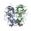
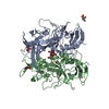
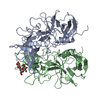
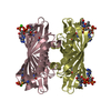
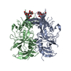
| Title | X-ray crystal structure of 4-hydroxybenzoyl CoA thioesterase mutant D17N complexed with 4-hydroxybenzoyl CoA | ||||||
 Components Components | 4-hydroxybenzoyl-CoA Thioesterase | ||||||
 Keywords Keywords | HYDROLASE / Thioesterase / hot dog fold / catalytic mechanism | ||||||
| Function / homology |  Function and homology information Function and homology information4-hydroxybenzoyl-CoA thioesterase / 4-hydroxybenzoyl-CoA thioesterase activity Similarity search - Function | ||||||
| Biological species |  Pseudomonas sp. CBS3 (bacteria) Pseudomonas sp. CBS3 (bacteria) | ||||||
| Method |  X-RAY DIFFRACTION / MOLECULAR REPLACEMENT / Resolution: 2.8 Å X-RAY DIFFRACTION / MOLECULAR REPLACEMENT / Resolution: 2.8 Å | ||||||
 Authors Authors | Thoden, J.B. / Holden, H.M. / Zhuang, Z. / Dunaway-Mariano, D. | ||||||
 Citation Citation | Journal: J.Biol.Chem. / Year: 2002 Title: X-ray crystallographic analyses of inhibitor and substrate complexes of wild-type and mutant 4-hydroxybenzoyl-CoA thioesterase. Authors: Thoden, J.B. / Holden, H.M. / Zhuang, Z. / Dunaway-Mariano, D. | ||||||
| History |
|
- Structure visualization
| Structure viewer | Molecule: MolmilJmol/JSmol |
|---|
- Downloads & links
-Download
| PDBx/mmCIF format | 1lo9.cif.gz | 43.9 KB | Display | PDBx/mmCIF format |
|---|---|---|---|---|
| PDB format | pdb1lo9.ent.gz | 29.4 KB | Display | PDB format |
| PDBx/mmJSON format | 1lo9.json.gz | Tree view | PDBx/mmJSON format | |
| Others |  Other downloads Other downloads |
-Validation report
| Arichive directory | https://data.pdbj.org/pub/pdb/validation_reports/lo/1lo9ftp://data.pdbj.org/pub/pdb/validation_reports/lo/1lo9 | HTTPS FTP |
|---|
-Related structure data
| Related structure data |  1lo7C  1lo8C  1bvqS C: citing same article ( S: Starting model for refinement |
|---|---|
| Similar structure data |


-Links
 PDBj
PDBj
- Assembly
| Deposited unit | 
| ||||||||||
|---|---|---|---|---|---|---|---|---|---|---|---|
| 1 | 
| ||||||||||
| Unit cell |
| ||||||||||
| Details | The biological assembly is a homotetramer. The single independent monomer in the asymmetric unit sits on 222 site symmetry. Matrices to generate the tetramer are as follows: (1) -1 0 0 0 -1 0 0 0 1 52.5 0.0 0.0 (2) -1 0 0 0 1 0 0 0 -1 52.5 0.0 0.0 (3) 1 0 0 0 -1 0 0 0 -1 0.0 0.0 0.0 |
-Components
| #1: Protein | Mass: 16139.466 Da / Num. of mol.: 1 / Mutation: D17N Source method: isolated from a genetically manipulated source Source: (gene. exp.) Pseudomonas sp. CBS3 (bacteria) / Strain: CBS-3 / Gene: 4HBT_PSESP / Plasmid: pET-3a / Production host: References: UniProt: P56653, 4-hydroxybenzoyl-CoA thioesterase |
|---|---|
| #2: Chemical | ChemComp-BCA /   Mass: 887.640 Da / Num. of mol.: 1 / Source method: obtained synthetically / Formula: C28H40N7O18P3S Mass: 887.640 Da / Num. of mol.: 1 / Source method: obtained synthetically / Formula: C28H40N7O18P3S |
| #3: Water | ChemComp-HOH /  Mass: 18.015 Da / Num. of mol.: 15 / Source method: isolated from a natural source / Formula: H2O Mass: 18.015 Da / Num. of mol.: 15 / Source method: isolated from a natural source / Formula: H2O |
-Experimental details
-Experiment
| Experiment | Method: X-RAY DIFFRACTION / Number of used crystals: 1 |
|---|
- Sample preparation
| Crystal | Density Matthews: 2.13 Å3/Da / Density % sol: 42.12 % | ||||||||||||||||||||||||||||||||||||||||||||||||||||||||
|---|---|---|---|---|---|---|---|---|---|---|---|---|---|---|---|---|---|---|---|---|---|---|---|---|---|---|---|---|---|---|---|---|---|---|---|---|---|---|---|---|---|---|---|---|---|---|---|---|---|---|---|---|---|---|---|---|---|
| Crystal grow | Temperature: 277 K / Method: batch / pH: 6 Details: PEG 5000-O-methylether, potassium chloride, MES, 4-hydroxybenzoyl CoA, pH 6.0, batch at 277K | ||||||||||||||||||||||||||||||||||||||||||||||||||||||||
| Crystal grow | *PLUS Temperature: 4 ℃ / pH: 7 / Method: sparse matrix screening | ||||||||||||||||||||||||||||||||||||||||||||||||||||||||
| Components of the solutions | *PLUS
|
-Data collection
| Diffraction | Mean temperature: 275 K |
|---|---|
| Diffraction source | Source: ROTATING ANODE / Type: RIGAKU RU200 / Wavelength: 1.5418 Å |
| Detector | Type: SIEMENS HI-STAR / Detector: AREA DETECTOR / Date: Oct 16, 2001 / Details: goebel mirrors |
| Radiation | Monochromator: goebel mirrors / Protocol: SINGLE WAVELENGTH / Monochromatic (M) / Laue (L): M / Scattering type: x-ray |
| Radiation wavelength | Wavelength: 1.5418 Å / Relative weight: 1 |
| Reflection | Resolution: 2.8→30 Å / Num. all: 3068 / Num. obs: 3068 / % possible obs: 85.1 % / Observed criterion σ(F): 0 / Observed criterion σ(I): 0 / Redundancy: 2.2 % / Rsym value: 0.078 / Net I/σ(I): 6.2 |
| Reflection shell | Resolution: 2.8→2.93 Å / Redundancy: 1.6 % / Mean I/σ(I) obs: 2.5 / Num. unique all: 349 / Rsym value: 0.285 / % possible all: 77.6 |
| Reflection | *PLUS Rmerge(I) obs: 0.078 |
| Reflection shell | *PLUS % possible obs: 77.6 % / Num. unique obs: 349 / Rmerge(I) obs: 0.285 |
- Processing
| Software |
| |||||||||||||||||||||||||
|---|---|---|---|---|---|---|---|---|---|---|---|---|---|---|---|---|---|---|---|---|---|---|---|---|---|---|
| Refinement | Method to determine structure: MOLECULAR REPLACEMENT Starting model: PDB ENTRY 1BVQ Resolution: 2.8→30 Å / σ(F): 0 / σ(I): 0 / Stereochemistry target values: Engh & Huber
| |||||||||||||||||||||||||
| Refinement step | Cycle: LAST / Resolution: 2.8→30 Å
| |||||||||||||||||||||||||
| Refine LS restraints |
| |||||||||||||||||||||||||
| Refinement | *PLUS Num. reflection obs: 2761 / Rfactor all: 0.148 / Rfactor Rfree: 0.247 / Rfactor Rwork: 0.148 | |||||||||||||||||||||||||
| Solvent computation | *PLUS | |||||||||||||||||||||||||
| Displacement parameters | *PLUS | |||||||||||||||||||||||||
| Refine LS restraints | *PLUS
|
