 Movie
Movie Controller

[English] 日本語
 Yorodumi
- PDB-1llq: Crystal Structure of Malic Enzyme from Ascaris suum Complexed wit... -
+ Open data

- Basic information
| Entry | Database: PDB / ID: 1llq | ||||||
|---|---|---|---|---|---|---|---|
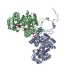
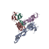
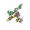
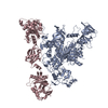
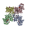
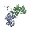
| Title | Crystal Structure of Malic Enzyme from Ascaris suum Complexed with Nicotinamide Adenine Dinucleotide | ||||||
 Components Components | NAD-dependent malic enzyme | ||||||
 Keywords Keywords | OXIDOREDUCTASE / Rossmann fold | ||||||
| Function / homology |  Function and homology information Function and homology informationmalate dehydrogenase (oxaloacetate-decarboxylating) / malate dehydrogenase (decarboxylating) (NAD+) activity / malate dehydrogenase (decarboxylating) (NADP+) activity / oxaloacetate decarboxylase activity / malate metabolic process / NAD binding / mitochondrial matrix / metal ion binding Similarity search - Function | ||||||
| Biological species |  Ascaris suum (pig roundworm) Ascaris suum (pig roundworm) | ||||||
| Method |  X-RAY DIFFRACTION / SYNCHROTRON / MOLECULAR REPLACEMENT / Resolution: 2.3 Å X-RAY DIFFRACTION / SYNCHROTRON / MOLECULAR REPLACEMENT / Resolution: 2.3 Å | ||||||
 Authors Authors | Coleman, D.E. / Jagannatha, G.S. / Goldsmith, E.J. / Cook, P.F. / Harris, B.G. | ||||||
 Citation Citation | Journal: Biochemistry / Year: 2002 Title: Crystal structure of the malic enzyme from Ascaris suum complexed with nicotinamide adenine dinucleotide at 2.3 A resolution. Authors: Coleman, D.E. / Rao, G.S. / Goldsmith, E.J. / Cook, P.F. / Harris, B.G. | ||||||
| History |
| ||||||
| Remark 999 | SEQUENCE The numbering system used for the coordinates corresponds to the numbering system used in ...SEQUENCE The numbering system used for the coordinates corresponds to the numbering system used in published mutational studies, where residue 1 (LYS) is the first residue present in the fully processed protein chain. A 12 residue mitochondrial transport sequence is present in the unprocessed protein, but is absent from processed protein and the protein present in the crystals. |
- Structure visualization
| Structure viewer | Molecule: MolmilJmol/JSmol |
|---|
- Downloads & links
-Download
| PDBx/mmCIF format | 1llq.cif.gz | 244.1 KB | Display | PDBx/mmCIF format |
|---|---|---|---|---|
| PDB format | pdb1llq.ent.gz | 197 KB | Display | PDB format |
| PDBx/mmJSON format | 1llq.json.gz | Tree view | PDBx/mmJSON format | |
| Others |  Other downloads Other downloads |
-Validation report
| Arichive directory | https://data.pdbj.org/pub/pdb/validation_reports/ll/1llqftp://data.pdbj.org/pub/pdb/validation_reports/ll/1llq | HTTPS FTP |
|---|
-Related structure data
| Related structure data | 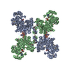 1qr6S S: Starting model for refinement |
|---|---|
| Similar structure data |
-Links
 PDBj
PDBj- Assembly
| Deposited unit | 
| ||||||||
|---|---|---|---|---|---|---|---|---|---|
| 1 | 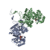
| ||||||||
| Unit cell |
| ||||||||
| Details | The Biological assembly is a tetramer generated from the dimer in the asymmetric unit by appling the following rotation and translation operators to the A and B subunits: -0.500000 0.866025 0.0 0.000 0.866025 0.500000 0.0 0.000 0.000000 0.000000 -1.0 149.230 |
-Components
| #1: Protein | Mass: 68556.406 Da / Num. of mol.: 2 / Source method: isolated from a natural source / Source: (natural) Ascaris suum (pig roundworm)References: UniProt: P27443, malate dehydrogenase (oxaloacetate-decarboxylating) #2: Chemical |   Mass: 663.425 Da / Num. of mol.: 2 / Source method: obtained synthetically / Formula: C21H27N7O14P2 / Comment: NAD*YM Mass: 663.425 Da / Num. of mol.: 2 / Source method: obtained synthetically / Formula: C21H27N7O14P2 / Comment: NAD*YM#3: Water | ChemComp-HOH / |  Mass: 18.015 Da / Num. of mol.: 74 / Source method: isolated from a natural source / Formula: H2O Mass: 18.015 Da / Num. of mol.: 74 / Source method: isolated from a natural source / Formula: H2O |
|---|
-Experimental details
-Experiment
| Experiment | Method: X-RAY DIFFRACTION / Number of used crystals: 1 |
|---|
- Sample preparation
| Crystal | Density Matthews: 2.68 Å3/Da / Density % sol: 54.09 % |
|---|---|
| Crystal grow | Temperature: 298 K / Method: vapor diffusion, hanging drop / pH: 7.5 Details: PEG 4000, tartronate, magnesium sulfate, TRIS, NAD, pH 7.5, VAPOR DIFFUSION, HANGING DROP, temperature 298.0K |
| Crystal grow | *PLUS Details: Clancy, L.L., (1992) J. Mol. Biol., 226, 565. |
-Data collection
| Diffraction | Mean temperature: 100 K |
|---|---|
| Diffraction source | Source: SYNCHROTRON / Site: CHESS  / Beamline: F1 / Wavelength: 0.947 Å / Beamline: F1 / Wavelength: 0.947 Å |
| Detector | Type: ADSC QUANTUM 4 / Detector: CCD / Date: Feb 5, 2000 |
| Radiation | Protocol: SINGLE WAVELENGTH / Monochromatic (M) / Laue (L): M / Scattering type: x-ray |
| Radiation wavelength | Wavelength: 0.947 Å / Relative weight: 1 |
| Reflection | Resolution: 2.3→25 Å / Num. all: 65007 / Num. obs: 65007 / % possible obs: 98.8 % / Observed criterion σ(F): 0 / Observed criterion σ(I): -3 / Redundancy: 4.7 % / Biso Wilson estimate: 38 Å2 / Rmerge(I) obs: 0.047 / Net I/σ(I): 30 |
| Reflection shell | Resolution: 2.3→2.37 Å / Redundancy: 4.5 % / Rmerge(I) obs: 0.224 / Mean I/σ(I) obs: 6.3 / Num. unique all: 5384 / % possible all: 99.3 |
| Reflection | *PLUS Num. obs: 62146 / % possible obs: 94.6 % / Num. measured all: 308683 / Rmerge(I) obs: 0.047 |
| Reflection shell | *PLUS Rmerge(I) obs: 0.224 |
- Processing
| Software |
| |||||||||||||||||||||||||
|---|---|---|---|---|---|---|---|---|---|---|---|---|---|---|---|---|---|---|---|---|---|---|---|---|---|---|
| Refinement | Method to determine structure: MOLECULAR REPLACEMENT Starting model: 1QR6 Resolution: 2.3→24.51 Å / Isotropic thermal model: isotropic / Cross valid method: THROUGHOUT / σ(F): 0 / Stereochemistry target values: Engh & Huber Details: Bulk solvent correction used throughout refinement.
| |||||||||||||||||||||||||
| Displacement parameters | Biso mean: 40.7 Å2 | |||||||||||||||||||||||||
| Refinement step | Cycle: LAST / Resolution: 2.3→24.51 Å
| |||||||||||||||||||||||||
| Refine LS restraints |
| |||||||||||||||||||||||||
| LS refinement shell | Resolution: 2.3→2.37 Å
| |||||||||||||||||||||||||
| Refinement | *PLUS Lowest resolution: 25 Å / % reflection Rfree: 5 % / Rfactor obs: 0.247 / Rfactor Rfree: 0.28 / Rfactor Rwork: 0.247 | |||||||||||||||||||||||||
| Solvent computation | *PLUS | |||||||||||||||||||||||||
| Displacement parameters | *PLUS | |||||||||||||||||||||||||
| LS refinement shell | *PLUS Highest resolution: 2.3 Å / Rfactor Rfree: 0.339 / Rfactor Rwork: 0.31 / Rfactor obs: 0.31 |
