 Movie
Movie Controller

[English] 日本語
 Yorodumi
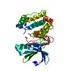
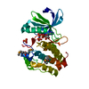
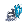
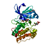
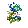
- PDB-1lln: 1.6A CRYSTAL STRUCTURE OF POKEWEED ANTIVIRAL PROTEIN-III (PAP-III... -
+ Open data

- Basic information
| Entry | Database: PDB / ID: 1lln | ||||||
|---|---|---|---|---|---|---|---|
| Title | 1.6A CRYSTAL STRUCTURE OF POKEWEED ANTIVIRAL PROTEIN-III (PAP-III) WITH METHYLATED LYSINES | ||||||
 Components Components | Antiviral Protein 3 | ||||||
 Keywords Keywords | HYDROLASE / POKEWEED ANTIVIRAL PROTEIN / RIBOSOME INACTIVATING PROTEIN / POLYNUCLEOTIDE:ADENOSINE | ||||||
| Function / homology |  Function and homology information Function and homology informationrRNA N-glycosylase / rRNA N-glycosylase activity / toxin activity / defense response to virus / negative regulation of translation Similarity search - Function | ||||||
| Biological species |  Phytolacca americana (American pokeweed) Phytolacca americana (American pokeweed) | ||||||
| Method |  X-RAY DIFFRACTION / SYNCHROTRON / MOLECULAR REPLACEMENT / Resolution: 1.6 Å X-RAY DIFFRACTION / SYNCHROTRON / MOLECULAR REPLACEMENT / Resolution: 1.6 Å | ||||||
 Authors Authors | Kurinov, I.V. / Uckun, F.M. | ||||||
 Citation Citation | Journal: Biochem.Pharm. / Year: 2003 Title: High resolution X-ray structure of potent anti-HIV pokeweed antiviral protein-III Authors: Kurinov, I.V. / Uckun, F.M. #1: Journal: Protein Sci. / Year: 1999Title: X-Ray Crystallographic Analysis of the Structural Basis for the Interactions of Pokeweed Antiviral Protein with its Active Site Inhibitor and Ribosomal RNA Substrate Analogs Authors: Kurinov, I.V. / Myers, D.E. / Irvin, J.D. / Uckun, F.M. | ||||||
| History |
|
- Structure visualization
| Structure viewer | Molecule: MolmilJmol/JSmol |
|---|
- Downloads & links
-Download
| PDBx/mmCIF format | 1lln.cif.gz | 69.6 KB | Display | PDBx/mmCIF format |
|---|---|---|---|---|
| PDB format | pdb1lln.ent.gz | 52 KB | Display | PDB format |
| PDBx/mmJSON format | 1lln.json.gz | Tree view | PDBx/mmJSON format | |
| Others |  Other downloads Other downloads |
-Validation report
| Arichive directory | https://data.pdbj.org/pub/pdb/validation_reports/ll/1llnftp://data.pdbj.org/pub/pdb/validation_reports/ll/1lln | HTTPS FTP |
|---|
-Related structure data
| Related structure data | |
|---|---|
| Similar structure data |


-Links
 PDBj
PDBj

- Assembly
| Deposited unit | 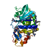
| ||||||||
|---|---|---|---|---|---|---|---|---|---|
| 1 |
| ||||||||
| Unit cell |
|
-Components
| #1: Protein | Mass: 29683.041 Da / Num. of mol.: 1 / Source method: isolated from a natural source / Details: late summer leaves / Source: (natural) Phytolacca americana (American pokeweed) / References: UniProt: Q40772, rRNA N-glycosylase |
|---|---|
| #2: Water | ChemComp-HOH /  Mass: 18.015 Da / Num. of mol.: 260 / Source method: isolated from a natural source / Formula: H2O Mass: 18.015 Da / Num. of mol.: 260 / Source method: isolated from a natural source / Formula: H2O |
| Has protein modification | Y |
-Experimental details
-Experiment
| Experiment | Method: X-RAY DIFFRACTION / Number of used crystals: 1 |
|---|
- Sample preparation
| Crystal | Density Matthews: 2.3 Å3/Da / Density % sol: 48.73 % | ||||||||||||||||||
|---|---|---|---|---|---|---|---|---|---|---|---|---|---|---|---|---|---|---|---|
| Crystal grow | Temperature: 290 K / Method: vapor diffusion, hanging drop / pH: 9.5 Details: 30% PEG400, pH 9.5, VAPOR DIFFUSION, HANGING DROP, temperature 290K | ||||||||||||||||||
| Crystal grow | *PLUS Temperature: 4 ℃ / pH: 8.5 / Method: vapor diffusion, hanging drop | ||||||||||||||||||
| Components of the solutions | *PLUS
|
-Data collection
| Diffraction | Mean temperature: 90 K |
|---|---|
| Diffraction source | Source: SYNCHROTRON / Site: SSRL  / Beamline: BL7-1 / Wavelength: 0.97 Å / Beamline: BL7-1 / Wavelength: 0.97 Å |
| Detector | Type: MARRESEARCH / Detector: IMAGE PLATE / Date: Mar 8, 2001 |
| Radiation | Protocol: SINGLE WAVELENGTH / Monochromatic (M) / Laue (L): M / Scattering type: x-ray |
| Radiation wavelength | Wavelength: 0.97 Å / Relative weight: 1 |
| Reflection | Resolution: 1.6→20 Å / Num. all: 38088 / Num. obs: 37018 / % possible obs: 97.3 % / Observed criterion σ(F): 0 / Observed criterion σ(I): 0 / Redundancy: 4.4 % / Rmerge(I) obs: 0.052 / Net I/σ(I): 23.9 |
| Reflection shell | Resolution: 1.6→1.66 Å / Rmerge(I) obs: 0.132 / % possible all: 96.9 |
| Reflection | *PLUS Highest resolution: 1.6 Å |
- Processing
| Software |
| ||||||||||||||||||||
|---|---|---|---|---|---|---|---|---|---|---|---|---|---|---|---|---|---|---|---|---|---|
| Refinement | Method to determine structure: MOLECULAR REPLACEMENT / Resolution: 1.6→7 Å / Data cutoff low absF: 0.0001 / Isotropic thermal model: Isotropic / Cross valid method: THROUGHOUT / σ(F): 2 / σ(I): 2 / Stereochemistry target values: Engh & Huber
| ||||||||||||||||||||
| Refinement step | Cycle: LAST / Resolution: 1.6→7 Å
| ||||||||||||||||||||
| Refine LS restraints |
| ||||||||||||||||||||
| Refinement | *PLUS Highest resolution: 1.6 Å / Rfactor Rwork: 0.203 | ||||||||||||||||||||
| Solvent computation | *PLUS | ||||||||||||||||||||
| Displacement parameters | *PLUS | ||||||||||||||||||||
| Refine LS restraints | *PLUS
|
