 Movie
Movie Controller

[English] 日本語
 Yorodumi
- PDB-1llf: Cholesterol Esterase (Candida Cylindracea) Crystal Structure at 1... -
+ Open data

- Basic information
| Entry | Database: PDB / ID: 1llf | |||||||||
|---|---|---|---|---|---|---|---|---|---|---|
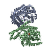
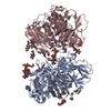
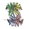
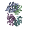
| Title | Cholesterol Esterase (Candida Cylindracea) Crystal Structure at 1.4A resolution | |||||||||
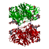
 Components Components | Lipase 3 | |||||||||
 Keywords Keywords | HYDROLASE / Candida cylindracea cholesterol esterase / sterol ester acylhydrolase | |||||||||
| Function / homology |  Function and homology information Function and homology informationtriacylglycerol lipase / triacylglycerol lipase activity / lipid catabolic process / cholesterol metabolic process / lipid metabolic process Similarity search - Function | |||||||||
| Biological species |  Candida cylindracea (fungus) Candida cylindracea (fungus) | |||||||||
| Method |  X-RAY DIFFRACTION / SYNCHROTRON / MOLECULAR REPLACEMENT / Resolution: 1.4 Å X-RAY DIFFRACTION / SYNCHROTRON / MOLECULAR REPLACEMENT / Resolution: 1.4 Å | |||||||||
 Authors Authors | Pletnev, V. / Addlagatta, A. / Wawrzak, Z. / Duax, W. | |||||||||
 Citation Citation | Journal: Acta Crystallogr.,Sect.D / Year: 2003 Title: Three-dimensional structure of homodimeric cholesterol esterase-ligand complex at 1.4 A resolution. Authors: Pletnev, V. / Addlagatta, A. / Wawrzak, Z. / Duax, W. #1: Journal: Structure / Year: 1995Title: Structure of uncomplexed and linoleate-bound Candida cylindracea cholesterol esterase Authors: Ghosh, D. / Wawrzak, Z. / Pletnev, V.Z. / Li, N. / Kaiser, R. / Pangborn, W. / Jornvall, H. / Erman, M. / Duax, W.L. | |||||||||
| History |
|
- Structure visualization
| Structure viewer | Molecule: MolmilJmol/JSmol |
|---|
- Downloads & links
-Download
| PDBx/mmCIF format | 1llf.cif.gz | 629.1 KB | Display | PDBx/mmCIF format |
|---|---|---|---|---|
| PDB format | pdb1llf.ent.gz | 525.2 KB | Display | PDB format |
| PDBx/mmJSON format | 1llf.json.gz | Tree view | PDBx/mmJSON format | |
| Others |  Other downloads Other downloads |
-Validation report
| Arichive directory | https://data.pdbj.org/pub/pdb/validation_reports/ll/1llfftp://data.pdbj.org/pub/pdb/validation_reports/ll/1llf | HTTPS FTP |
|---|
-Related structure data
| Related structure data |  1cleS S: Starting model for refinement |
|---|---|
| Similar structure data |


-Links
 PDBj
PDBj
- Assembly
| Deposited unit | 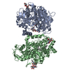
| ||||||||
|---|---|---|---|---|---|---|---|---|---|
| 1 |
| ||||||||
| Unit cell |
|
-Components
| #1: Protein | Mass: 57343.492 Da / Num. of mol.: 2 / Source method: isolated from a natural source / Source: (natural) Candida cylindracea (fungus)References: UniProt: p32947, UniProt: Q6S5M9*PLUS, triacylglycerol lipase #2: Polysaccharide | 2-acetamido-2-deoxy-beta-D-glucopyranose-(1-4)-2-acetamido-2-deoxy-beta-D-glucopyranose Source method: isolated from a genetically manipulated source #3: Chemical | 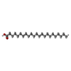  Mass: 354.610 Da / Num. of mol.: 2 / Source method: obtained synthetically / Formula: C23H46O2 Mass: 354.610 Da / Num. of mol.: 2 / Source method: obtained synthetically / Formula: C23H46O2#4: Water | ChemComp-HOH / |  Mass: 18.015 Da / Num. of mol.: 1078 / Source method: isolated from a natural source / Formula: H2O Mass: 18.015 Da / Num. of mol.: 1078 / Source method: isolated from a natural source / Formula: H2OHas protein modification | Y | |
|---|
-Experimental details
-Experiment
| Experiment | Method: X-RAY DIFFRACTION / Number of used crystals: 1 |
|---|
- Sample preparation
| Crystal | Density Matthews: 2.48 Å3/Da / Density % sol: 50.46 % | ||||||||||||||||||||||||
|---|---|---|---|---|---|---|---|---|---|---|---|---|---|---|---|---|---|---|---|---|---|---|---|---|---|
| Crystal grow | Temperature: 293 K / Method: vapor diffusion, hanging drop / pH: 7.3 Details: PEG 3350, potassium phosphate, detergent Thesit, pH 7.3, VAPOR DIFFUSION, HANGING DROP, temperature 293K | ||||||||||||||||||||||||
| Crystal grow | *PLUS | ||||||||||||||||||||||||
| Components of the solutions | *PLUS
|
-Data collection
| Diffraction | Mean temperature: 100 K |
|---|---|
| Diffraction source | Source: SYNCHROTRON / Site: APS  / Beamline: 5ID-B / Wavelength: 1 Å / Beamline: 5ID-B / Wavelength: 1 Å |
| Detector | Type: MARRESEARCH / Detector: CCD / Date: Feb 15, 2001 / Details: focusing mirrors |
| Radiation | Monochromator: double crystal Si monochromator / Protocol: SINGLE WAVELENGTH / Monochromatic (M) / Laue (L): M / Scattering type: x-ray |
| Radiation wavelength | Wavelength: 1 Å / Relative weight: 1 |
| Reflection | Resolution: 1.4→30 Å / Num. all: 206525 / Num. obs: 206525 / % possible obs: 90.2 % / Observed criterion σ(F): 0 / Observed criterion σ(I): 0 / Redundancy: 6.9 % / Rmerge(I) obs: 0.087 / Net I/σ(I): 14 |
| Reflection shell | Resolution: 1.4→1.5 Å / Rmerge(I) obs: 0.178 / Mean I/σ(I) obs: 7.8 / Num. unique all: 35868 / % possible all: 88 |
| Reflection | *PLUS Lowest resolution: 30 Å / Num. measured all: 1424531 |
| Reflection shell | *PLUS % possible obs: 88 % / Mean I/σ(I) obs: 7 |
- Processing
| Software |
| ||||||||||||||||||||||||||||||
|---|---|---|---|---|---|---|---|---|---|---|---|---|---|---|---|---|---|---|---|---|---|---|---|---|---|---|---|---|---|---|---|
| Refinement | Method to determine structure: MOLECULAR REPLACEMENT Starting model: PDB ENTRY 1CLE Resolution: 1.4→30 Å / Isotropic thermal model: anisotropic / Cross valid method: Free R / σ(F): 0 / σ(I): 0 / Stereochemistry target values: SHELDRICK Details: Konnert-Hendrickson conjugate gradient least squares algorithm
| ||||||||||||||||||||||||||||||
| Refine analyze | Luzzati coordinate error obs: 0.1 Å / Luzzati d res low obs: 5 Å | ||||||||||||||||||||||||||||||
| Refinement step | Cycle: LAST / Resolution: 1.4→30 Å
| ||||||||||||||||||||||||||||||
| Refine LS restraints |
| ||||||||||||||||||||||||||||||
| LS refinement shell |
| ||||||||||||||||||||||||||||||
| Software | *PLUS Name: SHELXL / Version: 97 / Classification: refinement | ||||||||||||||||||||||||||||||
| Refinement | *PLUS % reflection Rfree: 5 % | ||||||||||||||||||||||||||||||
| Solvent computation | *PLUS | ||||||||||||||||||||||||||||||
| Displacement parameters | *PLUS | ||||||||||||||||||||||||||||||
| Refine LS restraints | *PLUS
|
