 Movie
Movie Controller


+ Open data

- Basic information
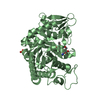
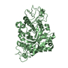
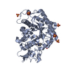
| Entry | Database: PDB / ID: 1ll4 | |||||||||
|---|---|---|---|---|---|---|---|---|---|---|
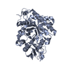
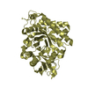
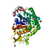
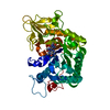
| Title | STRUCTURE OF C. IMMITIS CHITINASE 1 COMPLEXED WITH ALLOSAMIDIN | |||||||||
 Components Components | CHITINASE 1 | |||||||||
 Keywords Keywords | HYDROLASE / BETA-ALPHA BARREL / ENZYME-INHIBITOR COMPLEX | |||||||||
| Function / homology |  Function and homology information Function and homology informationendochitinase activity / chitinase / chitin catabolic process / chitin binding / polysaccharide catabolic process / extracellular region Similarity search - Function | |||||||||
| Biological species |  Coccidioides immitis (fungus) Coccidioides immitis (fungus) | |||||||||
| Method |  X-RAY DIFFRACTION / MOLECULAR REPLACEMENT / Resolution: 2.8 Å X-RAY DIFFRACTION / MOLECULAR REPLACEMENT / Resolution: 2.8 Å | |||||||||
 Authors Authors | Bortone, K. / Monzingo, A.F. / Ernst, S. / Robertus, J.D. | |||||||||
 Citation Citation | Journal: J.Mol.Biol. / Year: 2002 Title: THE STRUCTURE OF AN ALLOSAMIDIN COMPLEX WITH THE Coccidioides IMMITIS CHITINASE DEFINES A ROLE FOR A SECOND ACID RESIDUE IN SUBSTRATE-ASSISTED MECHANISM Authors: BORTONE, K. / MONZINGO, A.F. / ERNST, S. / ROBERTUS, J.D. | |||||||||
| History |
|
- Structure visualization
| Structure viewer | Molecule: MolmilJmol/JSmol |
|---|
- Downloads & links
-Download
| PDBx/mmCIF format | 1ll4.cif.gz | 312.1 KB | Display | PDBx/mmCIF format |
|---|---|---|---|---|
| PDB format | pdb1ll4.ent.gz | 252.8 KB | Display | PDB format |
| PDBx/mmJSON format | 1ll4.json.gz | Tree view | PDBx/mmJSON format | |
| Others |  Other downloads Other downloads |
-Validation report
| Summary document | 1ll4_validation.pdf.gz | 1.7 MB | Display | wwPDB validaton report |
|---|---|---|---|---|
| Full document | 1ll4_full_validation.pdf.gz | 1.7 MB | Display | |
| Data in XML | 1ll4_validation.xml.gz | 60.7 KB | Display | |
| Data in CIF | 1ll4_validation.cif.gz | 80.7 KB | Display | |
| Arichive directory | https://data.pdbj.org/pub/pdb/validation_reports/ll/1ll4ftp://data.pdbj.org/pub/pdb/validation_reports/ll/1ll4 | HTTPS FTP |
-Related structure data
| Related structure data |  1ll6C  1ll7C  1d2kS S: Starting model for refinement C: citing same article ( |
|---|---|
| Similar structure data |


-Links
 PDBj
PDBj- Assembly
| Deposited unit | 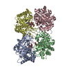
| ||||||||
|---|---|---|---|---|---|---|---|---|---|
| 1 | 
| ||||||||
| 2 | 
| ||||||||
| 3 | 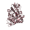
| ||||||||
| 4 | 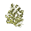
| ||||||||
| Unit cell |
|
-Components
| #1: Protein | Mass: 43714.812 Da / Num. of mol.: 4 / Fragment: RESIDUES 36-427 Source method: isolated from a genetically manipulated source Source: (gene. exp.) Coccidioides immitis (fungus) / Gene: CTS1 / Plasmid: pGEX-4T-3 / Production host:  #2: Polysaccharide | 2-acetamido-2-deoxy-beta-D-allopyranose-(1-4)-2-acetamido-2-deoxy-beta-D-allopyranose Source method: isolated from a genetically manipulated source #3: Chemical | ChemComp-AMI /   Mass: 216.234 Da / Num. of mol.: 4 / Source method: obtained synthetically / Formula: C9H16N2O4 Mass: 216.234 Da / Num. of mol.: 4 / Source method: obtained synthetically / Formula: C9H16N2O4#4: Water | ChemComp-HOH / |  Mass: 18.015 Da / Num. of mol.: 100 / Source method: isolated from a natural source / Formula: H2O Mass: 18.015 Da / Num. of mol.: 100 / Source method: isolated from a natural source / Formula: H2O |
|---|
-Experimental details
-Experiment
| Experiment | Method: X-RAY DIFFRACTION / Number of used crystals: 1 |
|---|
- Sample preparation
| Crystal | Density Matthews: 2.18 Å3/Da / Density % sol: 43.6 % | ||||||||||||||||||||||||||||||
|---|---|---|---|---|---|---|---|---|---|---|---|---|---|---|---|---|---|---|---|---|---|---|---|---|---|---|---|---|---|---|---|
| Crystal grow | Temperature: 298 K / Method: vapor diffusion, hanging drop / pH: 7.5 Details: PEG 4000, ISOPROPANOL, SODIUM HEPES, pH 7.5, VAPOR DIFFUSION, HANGING DROP, temperature 298K | ||||||||||||||||||||||||||||||
| Crystal grow | *PLUS pH: 4.6 / Method: vapor diffusion, sitting dropDetails: Hollis, T., (1998) Acta Crystallogr., Sect.D, 54, 1412. | ||||||||||||||||||||||||||||||
| Components of the solutions | *PLUS
|
-Data collection
| Diffraction | Mean temperature: 123 K |
|---|---|
| Diffraction source | Source: ROTATING ANODE / Type: RIGAKU RU200 / Wavelength: 1.5418 Å |
| Detector | Type: RIGAKU RAXIS IV / Detector: IMAGE PLATE / Date: Aug 19, 2000 |
| Radiation | Monochromator: DOUBLE FOCUSSING MIRRORS (NI & PT) + NI FILTER Protocol: SINGLE WAVELENGTH / Monochromatic (M) / Laue (L): M / Scattering type: x-ray |
| Radiation wavelength | Wavelength: 1.5418 Å / Relative weight: 1 |
| Reflection | Resolution: 2.8→50 Å / Num. all: 34859 / Num. obs: 34859 / % possible obs: 95.8 % / Observed criterion σ(I): 0 / Redundancy: 2.4 % / Rmerge(I) obs: 0.131 / Net I/σ(I): 5.6 |
| Reflection shell | Resolution: 2.8→2.9 Å / Redundancy: 2.3 % / Rmerge(I) obs: 0.369 / Mean I/σ(I) obs: 2.3 / Num. unique all: 3428 / % possible all: 93.2 |
| Reflection | *PLUS Rmerge(I) obs: 0.131 |
| Reflection shell | *PLUS Rmerge(I) obs: 0.369 |
- Processing
| Software |
| ||||||||||||||||||||
|---|---|---|---|---|---|---|---|---|---|---|---|---|---|---|---|---|---|---|---|---|---|
| Refinement | Method to determine structure: MOLECULAR REPLACEMENT Starting model: PDB ID 1D2K Resolution: 2.8→5 Å / Cross valid method: THROUGHOUT / σ(F): 2 / Stereochemistry target values: Engh & Huber
| ||||||||||||||||||||
| Refinement step | Cycle: LAST / Resolution: 2.8→5 Å
| ||||||||||||||||||||
| Refine LS restraints |
| ||||||||||||||||||||
| Refinement | *PLUS Rfactor Rfree: 0.258 / Rfactor Rwork: 0.197 | ||||||||||||||||||||
| Solvent computation | *PLUS | ||||||||||||||||||||
| Displacement parameters | *PLUS |
