 Movie
Movie Controller

[English] 日本語
 Yorodumi
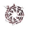
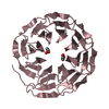
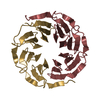
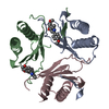
- PDB-1lkt: CRYSTAL STRUCTURE OF THE HEAD-BINDING DOMAIN OF PHAGE P22 TAILSPI... -
+ Open data

- Basic information
| Entry | Database: PDB / ID: 1lkt | ||||||
|---|---|---|---|---|---|---|---|
| Title | CRYSTAL STRUCTURE OF THE HEAD-BINDING DOMAIN OF PHAGE P22 TAILSPIKE PROTEIN | ||||||
 Components Components | TAILSPIKE PROTEIN | ||||||
 Keywords Keywords | VIRAL PROTEIN / VIRUS PROTEIN / SALMONELLA PHAGE P22 / TELLUROMETHIONINE / LATE PROTEIN | ||||||
| Function / homology |  Function and homology information Function and homology informationendo-1,3-alpha-L-rhamnosidase activity / symbiont entry into host cell via disruption of host cell envelope lipopolysaccharide / virus tail, fiber / symbiont entry into host cell via disruption of host cell envelope / symbiont entry into host / Hydrolases; Glycosylases; Glycosidases, i.e. enzymes that hydrolyse O- and S-glycosyl compounds / adhesion receptor-mediated virion attachment to host cell / virion attachment to host cell Similarity search - Function | ||||||
| Biological species |  Enterobacteria phage P22 (virus) Enterobacteria phage P22 (virus) | ||||||
| Method |  X-RAY DIFFRACTION / MIR / Resolution: 2.6 Å X-RAY DIFFRACTION / MIR / Resolution: 2.6 Å | ||||||
 Authors Authors | Steinbacher, S. | ||||||
 Citation Citation | Journal: J.Mol.Biol. / Year: 1997 Title: Phage P22 tailspike protein: crystal structure of the head-binding domain at 2.3 A, fully refined structure of the endorhamnosidase at 1.56 A resolution, and the molecular basis of O-antigen ...Title: Phage P22 tailspike protein: crystal structure of the head-binding domain at 2.3 A, fully refined structure of the endorhamnosidase at 1.56 A resolution, and the molecular basis of O-antigen recognition and cleavage. Authors: Steinbacher, S. / Miller, S. / Baxa, U. / Budisa, N. / Weintraub, A. / Seckler, R. / Huber, R. | ||||||
| History |
|
- Structure visualization
| Structure viewer | Molecule: MolmilJmol/JSmol |
|---|
- Downloads & links
-Download
| PDBx/mmCIF format | 1lkt.cif.gz | 171.7 KB | Display | PDBx/mmCIF format |
|---|---|---|---|---|
| PDB format | pdb1lkt.ent.gz | 139.6 KB | Display | PDB format |
| PDBx/mmJSON format | 1lkt.json.gz | Tree view | PDBx/mmJSON format | |
| Others |  Other downloads Other downloads |
-Validation report
| Summary document | 1lkt_validation.pdf.gz | 401.8 KB | Display | wwPDB validaton report |
|---|---|---|---|---|
| Full document | 1lkt_full_validation.pdf.gz | 416.5 KB | Display | |
| Data in XML | 1lkt_validation.xml.gz | 13.5 KB | Display | |
| Data in CIF | 1lkt_validation.cif.gz | 23.9 KB | Display | |
| Arichive directory | https://data.pdbj.org/pub/pdb/validation_reports/lk/1lktftp://data.pdbj.org/pub/pdb/validation_reports/lk/1lkt | HTTPS FTP |
-Related structure data
| Similar structure data |
|---|


-Links
 PDBj
PDBj- Assembly
| Deposited unit | 
| ||||||||||||||||||||||||
|---|---|---|---|---|---|---|---|---|---|---|---|---|---|---|---|---|---|---|---|---|---|---|---|---|---|
| 1 | 
| ||||||||||||||||||||||||
| 2 | 
| ||||||||||||||||||||||||
| Unit cell |
| ||||||||||||||||||||||||
| Noncrystallographic symmetry (NCS) | NCS oper:
|
-Components
| #1: Protein | Mass: 11307.753 Da / Num. of mol.: 6 / Fragment: HEAD-BINDING DOMAIN Source method: isolated from a genetically manipulated source Source: (gene. exp.) Enterobacteria phage P22 (virus) / Genus: P22-like viruses / Cell line: BL21 / Organ: TAIL / Plasmid: BL21 / Species (production host): Escherichia coli / Production host:  #2: Water | ChemComp-HOH / |  Mass: 18.015 Da / Num. of mol.: 432 / Source method: isolated from a natural source / Formula: H2O Mass: 18.015 Da / Num. of mol.: 432 / Source method: isolated from a natural source / Formula: H2O |
|---|
-Experimental details
-Experiment
| Experiment | Method: X-RAY DIFFRACTION / Number of used crystals: 1 |
|---|
- Sample preparation
| Crystal | Density Matthews: 2.05 Å3/Da / Density % sol: 51.89 % | ||||||||||||||||||||||||
|---|---|---|---|---|---|---|---|---|---|---|---|---|---|---|---|---|---|---|---|---|---|---|---|---|---|
| Crystal grow | pH: 6.6 / Details: 20% PEG 8K, 0.2 M MGCL2, 0.1 M BIS-TRIS, PH 6.6 | ||||||||||||||||||||||||
| Crystal grow | *PLUS Method: vapor diffusion | ||||||||||||||||||||||||
| Components of the solutions | *PLUS
|
-Data collection
| Diffraction | Mean temperature: 300 K |
|---|---|
| Diffraction source | Source: ROTATING ANODE / Type: RIGAKU RUH2R / Wavelength: 1.5418 |
| Detector | Type: MARRESEARCH / Detector: IMAGE PLATE / Date: Apr 1, 1996 |
| Radiation | Monochromator: GRAPHITE(002) / Monochromatic (M) / Laue (L): M / Scattering type: x-ray |
| Radiation wavelength | Wavelength: 1.5418 Å / Relative weight: 1 |
| Reflection | Resolution: 2.6→25 Å / Num. obs: 20822 / % possible obs: 96.5 % / Observed criterion σ(I): 2 / Redundancy: 2.3 % / Rmerge(I) obs: 0.076 |
| Reflection shell | Resolution: 2.6→2.65 Å / Rmerge(I) obs: 0.298 / % possible all: 72.2 |
| Reflection | *PLUS Num. measured all: 48766 |
| Reflection shell | *PLUS % possible obs: 72.2 % |
- Processing
| Software |
| ||||||||||||||||||||||||||||||||||||||||||||||||||||||||||||
|---|---|---|---|---|---|---|---|---|---|---|---|---|---|---|---|---|---|---|---|---|---|---|---|---|---|---|---|---|---|---|---|---|---|---|---|---|---|---|---|---|---|---|---|---|---|---|---|---|---|---|---|---|---|---|---|---|---|---|---|---|---|
| Refinement | Method to determine structure: MIR / Resolution: 2.6→8 Å / σ(F): 2
| ||||||||||||||||||||||||||||||||||||||||||||||||||||||||||||
| Refinement step | Cycle: LAST / Resolution: 2.6→8 Å
| ||||||||||||||||||||||||||||||||||||||||||||||||||||||||||||
| Refine LS restraints |
| ||||||||||||||||||||||||||||||||||||||||||||||||||||||||||||
| Software | *PLUS Name: X-PLOR / Classification: refinement | ||||||||||||||||||||||||||||||||||||||||||||||||||||||||||||
| Refinement | *PLUS Num. reflection obs: 18825 / Rfactor obs: 0.186 | ||||||||||||||||||||||||||||||||||||||||||||||||||||||||||||
| Solvent computation | *PLUS | ||||||||||||||||||||||||||||||||||||||||||||||||||||||||||||
| Displacement parameters | *PLUS | ||||||||||||||||||||||||||||||||||||||||||||||||||||||||||||
| Refine LS restraints | *PLUS
|
