 Movie
Movie Controller

[English] 日本語
 Yorodumi
- PDB-1lk6: Structure of dimeric antithrombin complexed with a P14-P9 reactiv... -
+ Open data

- Basic information
| Entry | Database: PDB / ID: 1lk6 | |||||||||
|---|---|---|---|---|---|---|---|---|---|---|
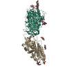
| Title | Structure of dimeric antithrombin complexed with a P14-P9 reactive loop peptide and an exogenous tripeptide | |||||||||
 Components Components |
| |||||||||
 Keywords Keywords | HYDROLASE/HYDROLASE INHIBITOR / Loop-sheet polymer / beta-barrel / BLOOD CLOTTING / HYDROLASE-HYDROLASE INHIBITOR COMPLEX | |||||||||
| Function / homology |  Function and homology information Function and homology informationregulation of blood coagulation / : / : / Post-translational protein phosphorylation / serine-type endopeptidase inhibitor activity / Regulation of Insulin-like Growth Factor (IGF) transport and uptake by Insulin-like Growth Factor Binding Proteins (IGFBPs) / blood coagulation / heparin binding / extracellular matrix / protease binding ...regulation of blood coagulation / : / : / Post-translational protein phosphorylation / serine-type endopeptidase inhibitor activity / Regulation of Insulin-like Growth Factor (IGF) transport and uptake by Insulin-like Growth Factor Binding Proteins (IGFBPs) / blood coagulation / heparin binding / extracellular matrix / protease binding / blood microparticle / endoplasmic reticulum lumen / : / extracellular exosome / extracellular region / identical protein binding / plasma membrane Similarity search - Function | |||||||||
| Biological species |  Homo sapiens (human) Homo sapiens (human) | |||||||||
| Method |  X-RAY DIFFRACTION / SYNCHROTRON / MOLECULAR REPLACEMENT / Resolution: 2.8 Å X-RAY DIFFRACTION / SYNCHROTRON / MOLECULAR REPLACEMENT / Resolution: 2.8 Å | |||||||||
 Authors Authors | Zhou, A. / Huntington, J.A. / Lomas, D.A. / Carrell, R.W. / Stein, P.E. | |||||||||
 Citation Citation | Journal: J.Biol.Chem. / Year: 2003 Title: Serpin Polymerization Is Prevented by a Hydrogen Bond Network That Is Centered on His-334 and Stabilized by Glycerol Authors: Zhou, A. / Stein, P.E. / Huntington, J.A. / Carrell, R.W. | |||||||||
| History |
|
- Structure visualization
| Structure viewer | Molecule: MolmilJmol/JSmol |
|---|
- Downloads & links
-Download
| PDBx/mmCIF format | 1lk6.cif.gz | 163.4 KB | Display | PDBx/mmCIF format |
|---|---|---|---|---|
| PDB format | pdb1lk6.ent.gz | 131 KB | Display | PDB format |
| PDBx/mmJSON format | 1lk6.json.gz | Tree view | PDBx/mmJSON format | |
| Others |  Other downloads Other downloads |
-Validation report
| Arichive directory | https://data.pdbj.org/pub/pdb/validation_reports/lk/1lk6ftp://data.pdbj.org/pub/pdb/validation_reports/lk/1lk6 | HTTPS FTP |
|---|
-Related structure data
| Related structure data |  1jvqS S: Starting model for refinement |
|---|---|
| Similar structure data |


-Links
 PDBj
PDBj


- Assembly
| Deposited unit | 
| ||||||||||
|---|---|---|---|---|---|---|---|---|---|---|---|
| 1 |
| ||||||||||
| Unit cell |
|
-Components
-Protein , 1 types, 2 molecules LI
| #1: Protein | Mass: 49101.016 Da / Num. of mol.: 2 / Source method: isolated from a natural source / Source: (natural) Homo sapiens (human) / Tissue: blood / Tissue fraction: plasma / References: UniProt: P01008 |
|---|
-Protein/peptide , 2 types, 2 molecules CD
| #2: Protein/peptide | Mass: 560.556 Da / Num. of mol.: 1 / Source method: obtained synthetically Details: This sequence ocurs naturally in human antithrombin |
|---|---|
| #3: Protein/peptide | Mass: 437.553 Da / Num. of mol.: 1 / Source method: obtained synthetically |
-Sugars , 2 types, 8 molecules 

| #4: Sugar | ChemComp-NDG /  Type: D-saccharide, alpha linking / Mass: 221.208 Da / Num. of mol.: 6 Type: D-saccharide, alpha linking / Mass: 221.208 Da / Num. of mol.: 6Source method: isolated from a genetically manipulated source Formula: C8H15NO6 #5: Sugar |  Type: D-saccharide, beta linking / Mass: 221.208 Da / Num. of mol.: 2 Type: D-saccharide, beta linking / Mass: 221.208 Da / Num. of mol.: 2Source method: isolated from a genetically manipulated source Formula: C8H15NO6 |
|---|
-Non-polymers , 2 types, 35 molecules 

| #6: Chemical | ChemComp-GOL /  Mass: 92.094 Da / Num. of mol.: 1 / Source method: obtained synthetically / Formula: C3H8O3 Mass: 92.094 Da / Num. of mol.: 1 / Source method: obtained synthetically / Formula: C3H8O3 |
|---|---|
| #7: Water | ChemComp-HOH / Mass: 18.015 Da / Num. of mol.: 34 / Source method: isolated from a natural source / Formula: H2O |
-Details
| Has protein modification | Y |
|---|
-Experimental details
-Experiment
| Experiment | Method: X-RAY DIFFRACTION / Number of used crystals: 1 |
|---|
- Sample preparation
| Crystal | Density Matthews: 3.07 Å3/Da / Density % sol: 59.6 % | ||||||||||||||||||||||||||||||||||||||||||
|---|---|---|---|---|---|---|---|---|---|---|---|---|---|---|---|---|---|---|---|---|---|---|---|---|---|---|---|---|---|---|---|---|---|---|---|---|---|---|---|---|---|---|---|
| Crystal grow | Temperature: 298 K / Method: vapor diffusion, hanging drop / pH: 6.8 Details: PEG 4000, sodium cacodylate, ammonium fluoride, glycerol, pH 6.8, VAPOR DIFFUSION, HANGING DROP at 298K | ||||||||||||||||||||||||||||||||||||||||||
| Crystal grow | *PLUS Method: vapor diffusion, hanging drop | ||||||||||||||||||||||||||||||||||||||||||
| Components of the solutions | *PLUS
|
-Data collection
| Diffraction | Mean temperature: 100 K |
|---|---|
| Diffraction source | Source: SYNCHROTRON / Site: SRS  / Beamline: PX14.2 / Wavelength: 0.978 Å / Beamline: PX14.2 / Wavelength: 0.978 Å |
| Detector | Type: ADSC QUANTUM 4 / Detector: CCD / Date: Jan 30, 2002 / Details: mirrors |
| Radiation | Monochromator: cooled liquid gallium / Protocol: SINGLE WAVELENGTH / Monochromatic (M) / Laue (L): M / Scattering type: x-ray |
| Radiation wavelength | Wavelength: 0.978 Å / Relative weight: 1 |
| Reflection | Resolution: 2.8→37.58 Å / Num. all: 27398 / Num. obs: 27376 / % possible obs: 96.7 % / Observed criterion σ(I): -3.7 / Redundancy: 3.3 % / Biso Wilson estimate: 51.2 Å2 / Rmerge(I) obs: 0.104 / Rsym value: 0.087 / Net I/σ(I): 7.5 |
| Reflection shell | Resolution: 2.8→2.95 Å / Redundancy: 2.5 % / Rmerge(I) obs: 0.551 / Mean I/σ(I) obs: 1.7 / Num. unique all: 3351 / Rsym value: 0.431 / % possible all: 81.5 |
| Reflection | *PLUS Highest resolution: 2.8 Å / Num. measured all: 91051 |
| Reflection shell | *PLUS Lowest resolution: 2.93 Å / % possible obs: 81.5 % |
- Processing
| Software |
| |||||||||||||||||||||||||||||||||||||||||||||||||
|---|---|---|---|---|---|---|---|---|---|---|---|---|---|---|---|---|---|---|---|---|---|---|---|---|---|---|---|---|---|---|---|---|---|---|---|---|---|---|---|---|---|---|---|---|---|---|---|---|---|---|
| Refinement | Method to determine structure: MOLECULAR REPLACEMENT Starting model: Dimeric antithrombin complexed with a P14-P8 reactive loop peptide and an exogenous tetrapeptide (1jvq). Both peptides omitted in starting model Resolution: 2.8→34.03 Å / Isotropic thermal model: Restrained / Cross valid method: THROUGHOUT / σ(F): 0 / Stereochemistry target values: Engh & Huber / Details: Maximum likelihood target
| |||||||||||||||||||||||||||||||||||||||||||||||||
| Displacement parameters | Biso mean: 55.26 Å2
| |||||||||||||||||||||||||||||||||||||||||||||||||
| Refine analyze |
| |||||||||||||||||||||||||||||||||||||||||||||||||
| Refinement step | Cycle: LAST / Resolution: 2.8→34.03 Å
| |||||||||||||||||||||||||||||||||||||||||||||||||
| Refine LS restraints |
| |||||||||||||||||||||||||||||||||||||||||||||||||
| LS refinement shell | Refine-ID: X-RAY DIFFRACTION / Total num. of bins used: 6
| |||||||||||||||||||||||||||||||||||||||||||||||||
| Refinement | *PLUS Highest resolution: 2.8 Å / Lowest resolution: 34 Å | |||||||||||||||||||||||||||||||||||||||||||||||||
| Solvent computation | *PLUS | |||||||||||||||||||||||||||||||||||||||||||||||||
| Displacement parameters | *PLUS | |||||||||||||||||||||||||||||||||||||||||||||||||
| Refine LS restraints | *PLUS
|
