 Movie
Movie Controller

[English] 日本語
 Yorodumi
- PDB-1l7f: Crystal structure of influenza virus neuraminidase in complex wit... -
+ Open data

- Basic information
| Entry | Database: PDB / ID: 1l7f | |||||||||
|---|---|---|---|---|---|---|---|---|---|---|
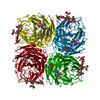
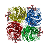
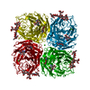
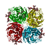
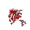
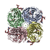
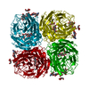
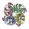
| Title | Crystal structure of influenza virus neuraminidase in complex with BCX-1812 | |||||||||
 Components Components | neuraminidase | |||||||||
 Keywords Keywords | HYDROLASE / N9 neuraminidase / influenza / glycosylated protein / BCX-1812 | |||||||||
| Function / homology |  Function and homology information Function and homology informationexo-alpha-sialidase / exo-alpha-sialidase activity / viral budding from plasma membrane / carbohydrate metabolic process / host cell plasma membrane / virion membrane / membrane / metal ion binding Similarity search - Function | |||||||||
| Biological species |   Influenza A virus Influenza A virus | |||||||||
| Method |  X-RAY DIFFRACTION / MOLECULAR REPLACEMENT / Resolution: 1.8 Å X-RAY DIFFRACTION / MOLECULAR REPLACEMENT / Resolution: 1.8 Å | |||||||||
 Authors Authors | Smith, B.J. / McKimm-Breshkin, J.L. / McDonald, M. / Fernley, R.T. / Varghese, J.N. / Colman, P.M. | |||||||||
 Citation Citation | Journal: J.Med.Chem. / Year: 2002 Title: Structural studies of the resistance of influenza virus neuramindase to inhibitors. Authors: Smith, B.J. / McKimm-Breshkin, J.L. / McDonald, M. / Fernley, R.T. / Varghese, J.N. / Colman, P.M. #1: Journal: Protein Sci. / Year: 1995Title: Three-dimensional structure of the complex of 4-guanidino-Neu5Ac2en and influenza virus neuraminidase Authors: Varghese, J.N. / Epa, V.C. / Colman, P.M. #2: Journal: Structure / Year: 1998Title: Drug design against a shifting target: a structural basis for resistance to inhibitors in a variant of influenza virus neuraminidase Authors: Varghese, J.N. / Smith, P.W. / Sollis, S.L. / Blick, T.J. / Sahasrabudhe, A. / McKimm-Breschkin, J.L. / Colman, P.M. | |||||||||
| History |
|
- Structure visualization
| Structure viewer | Molecule: MolmilJmol/JSmol |
|---|
- Downloads & links
-Download
| PDBx/mmCIF format | 1l7f.cif.gz | 113.5 KB | Display | PDBx/mmCIF format |
|---|---|---|---|---|
| PDB format | pdb1l7f.ent.gz | 84.6 KB | Display | PDB format |
| PDBx/mmJSON format | 1l7f.json.gz | Tree view | PDBx/mmJSON format | |
| Others |  Other downloads Other downloads |
-Validation report
| Arichive directory | https://data.pdbj.org/pub/pdb/validation_reports/l7/1l7fftp://data.pdbj.org/pub/pdb/validation_reports/l7/1l7f | HTTPS FTP |
|---|
-Related structure data


-Links
 PDBj
PDBj

- Assembly
| Deposited unit | 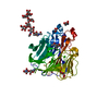
| ||||||||||||
|---|---|---|---|---|---|---|---|---|---|---|---|---|---|
| 1 | 
| ||||||||||||
| Unit cell |
| ||||||||||||
| Components on special symmetry positions |
|
-Components
-Protein , 1 types, 1 molecules A
| #1: Protein | Mass: 43723.770 Da / Num. of mol.: 1 Fragment: integral membrane protein, membrane stalk cleaved by pronase releasing fully active residues 82-468 Source method: isolated from a natural source / Source: (natural) Influenza A virus / Genus: Influenzavirus A / Strain: A/NWS/Tern/Australia/G70C/75 / References: UniProt: P03472, exo-alpha-sialidase |
|---|
-Sugars , 3 types, 3 molecules 
| #2: Polysaccharide | alpha-D-mannopyranose-(1-2)-alpha-D-mannopyranose-(1-2)-alpha-D-mannopyranose-(1-3)-[alpha-D- ...alpha-D-mannopyranose-(1-2)-alpha-D-mannopyranose-(1-2)-alpha-D-mannopyranose-(1-3)-[alpha-D-mannopyranose-(1-6)]beta-D-mannopyranose-(1-4)-2-acetamido-2-deoxy-beta-D-glucopyranose-(1-4)-2-acetamido-2-deoxy-beta-D-glucopyranose Source method: isolated from a genetically manipulated source |
|---|---|
| #3: Polysaccharide | 2-acetamido-2-deoxy-beta-D-glucopyranose-(1-4)-2-acetamido-2-deoxy-beta-D-glucopyranose Source method: isolated from a genetically manipulated source |
| #4: Sugar | ChemComp-NAG /  Type: D-saccharide, beta linking / Mass: 221.208 Da / Num. of mol.: 1 Type: D-saccharide, beta linking / Mass: 221.208 Da / Num. of mol.: 1Source method: isolated from a genetically manipulated source Formula: C8H15NO6 |
-Non-polymers , 4 types, 648 molecules 


| #5: Chemical |  Mass: 40.078 Da / Num. of mol.: 2 / Source method: obtained synthetically / Formula: Ca Mass: 40.078 Da / Num. of mol.: 2 / Source method: obtained synthetically / Formula: Ca#6: Chemical | ChemComp-BCZ / |  Mass: 328.407 Da / Num. of mol.: 1 / Source method: obtained synthetically / Formula: C15H28N4O4 / Comment: antivirus, inhibitor*YM Mass: 328.407 Da / Num. of mol.: 1 / Source method: obtained synthetically / Formula: C15H28N4O4 / Comment: antivirus, inhibitor*YM#7: Chemical |  Mass: 92.094 Da / Num. of mol.: 3 / Source method: obtained synthetically / Formula: C3H8O3 Mass: 92.094 Da / Num. of mol.: 3 / Source method: obtained synthetically / Formula: C3H8O3#8: Water | ChemComp-HOH / | Mass: 18.015 Da / Num. of mol.: 642 / Source method: isolated from a natural source / Formula: H2O |
|---|
-Details
| Has protein modification | Y |
|---|
-Experimental details
-Experiment
| Experiment | Method: X-RAY DIFFRACTION / Number of used crystals: 1 |
|---|
- Sample preparation
| Crystal | Density Matthews: 2.83 Å3/Da / Density % sol: 56.53 % | ||||||||||||||||||||
|---|---|---|---|---|---|---|---|---|---|---|---|---|---|---|---|---|---|---|---|---|---|
| Crystal grow | Temperature: 293 K / Method: vapor diffusion, hanging drop / pH: 5.9 Details: phosphate, pH 5.9, VAPOR DIFFUSION, HANGING DROP, temperature 293K | ||||||||||||||||||||
| Crystal grow | *PLUS pH: 6.6 / Method: vapor diffusion / Details: Laver, W.G., (1984) Virology, 137, 314. | ||||||||||||||||||||
| Components of the solutions | *PLUS
|
-Data collection
| Diffraction | Mean temperature: 113 K |
|---|---|
| Diffraction source | Source: ROTATING ANODE / Type: MACSCIENCE / Wavelength: 1.5418 Å |
| Detector | Type: RIGAKU RAXIS IV / Detector: IMAGE PLATE / Details: elliptical glass monocapillary |
| Radiation | Monochromator: Ni FILTER / Protocol: SINGLE WAVELENGTH / Monochromatic (M) / Laue (L): M / Scattering type: x-ray |
| Radiation wavelength | Wavelength: 1.5418 Å / Relative weight: 1 |
| Reflection | Resolution: 1.8→20 Å / Num. obs: 46825 / % possible obs: 100 % / Observed criterion σ(I): -3 / Rmerge(I) obs: 0.15 / Net I/σ(I): 28.5 |
| Reflection shell | Resolution: 1.8→1.84 Å / Rmerge(I) obs: 0.58 / Mean I/σ(I) obs: 3.2 / % possible all: 100 |
| Reflection | *PLUS Num. measured all: 453337 |
| Reflection shell | *PLUS % possible obs: 100 % |
- Processing
| Software |
| ||||||||||||||||||||
|---|---|---|---|---|---|---|---|---|---|---|---|---|---|---|---|---|---|---|---|---|---|
| Refinement | Method to determine structure: MOLECULAR REPLACEMENT / Resolution: 1.8→6 Å / σ(F): 0 / σ(I): 0 / Stereochemistry target values: Engh & Huber
| ||||||||||||||||||||
| Refinement step | Cycle: LAST / Resolution: 1.8→6 Å
| ||||||||||||||||||||
| Refine LS restraints |
| ||||||||||||||||||||
| LS refinement shell | Resolution: 1.8→1.865 Å
| ||||||||||||||||||||
| Refinement | *PLUS Rfactor obs: 0.153 | ||||||||||||||||||||
| Solvent computation | *PLUS | ||||||||||||||||||||
| Displacement parameters | *PLUS | ||||||||||||||||||||
| Refine LS restraints | *PLUS Type: p_planar_d / Dev ideal: 0.023 |
