 ムービー
ムービー コントローラー


+ データを開く

- 基本情報
基本情報
| 登録情報 | データベース: PDB / ID: 1l6e | ||||||
|---|---|---|---|---|---|---|---|
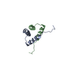
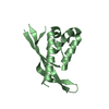
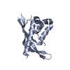
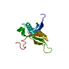
| タイトル | Solution structure of the docking and dimerization domain of protein kinase A II-alpha (RIIalpha D/D). Alternatively called the N-terminal dimerization domain of the regulatory subunit of protein kinase A. | ||||||
 要素 要素 | cAMP-dependent protein kinase Type II-alpha regulatory chain | ||||||
 キーワード キーワード | TRANSFERASE / Four-helix bundle / helix-loop-helix / regulatory subunit / dimerization / docking / anchoring | ||||||
| 機能・相同性 |  機能・相同性情報 機能・相同性情報PKA activation in glucagon signalling / CREB1 phosphorylation through the activation of Adenylate Cyclase / DARPP-32 events / PKA activation / Vasopressin regulates renal water homeostasis via Aquaporins / GPER1 signaling / Hedgehog 'off' state / Factors involved in megakaryocyte development and platelet production / cAMP-dependent protein kinase regulator activity / High laminar flow shear stress activates signaling by PIEZO1 and PECAM1:CDH5:KDR in endothelial cells ...PKA activation in glucagon signalling / CREB1 phosphorylation through the activation of Adenylate Cyclase / DARPP-32 events / PKA activation / Vasopressin regulates renal water homeostasis via Aquaporins / GPER1 signaling / Hedgehog 'off' state / Factors involved in megakaryocyte development and platelet production / cAMP-dependent protein kinase regulator activity / High laminar flow shear stress activates signaling by PIEZO1 and PECAM1:CDH5:KDR in endothelial cells / nucleotide-activated protein kinase complex / cAMP-dependent protein kinase inhibitor activity / beta-2 adrenergic receptor binding / cAMP-dependent protein kinase complex / small molecule binding / protein kinase A catalytic subunit binding / plasma membrane raft / cAMP binding / negative regulation of cAMP/PKA signal transduction / T-tubule / modulation of chemical synaptic transmission / adenylate cyclase-activating G protein-coupled receptor signaling pathway / protein domain specific binding / ubiquitin protein ligase binding / centrosome / synapse / protein-containing complex binding / perinuclear region of cytoplasm / glutamatergic synapse / protein-containing complex / identical protein binding / plasma membrane / cytoplasm / cytosol 類似検索 - 分子機能 | ||||||
| 生物種 |  | ||||||
| 手法 | 溶液NMR / Hybrid distance geometry-dynamical simulated annealing, refinement protocol for monomer structure determination, with 457 NOE-derived distance restraints (185 intra-residue, i-j=0; 136 sequential, |i-j|=1; 95 medium range, 1<|i-j|<5; 41 long range, |i-j|>4), 19 distance restraints representing hydrogen bonds (entered as 2 distances each), 25 phi-, 5 chi1-torsion angle restraints. Molecular dynamical simulated annealing protocol for dimer structure determination, using 505 NOE-derived distance restraints (185 intra-residue, i-j=0; 136 sequential, |i-j|=1; 95 medium range, 1<|i-j|<5; 25 long range, |i-j|>4; 38 inter-molecular; 26 ambiguous), 19 distance restraints representing hydrogen bonds (entered as 2 distances each), 25 phi-, 5 chi1-torsion angle restraints. | ||||||
 データ登録者 データ登録者 | Morikis, D. / Roy, M. / Newlon, M.G. / Scott, J.D. / Jennings, P.A. | ||||||
 引用 引用 | ジャーナル: Eur.J.Biochem. / 年: 2002 タイトル: Electrostatic properties of the structure of the docking and dimerization domain of protein kinase A IIalpha 著者: Morikis, D. / Roy, M. / Newlon, M.G. / Scott, J.D. / Jennings, P.A. #1: ジャーナル: Nat.Struct.Biol. / 年: 1999タイトル: The molecular basis for protein kinase A anchoring revealed by solution NMR. 著者: Newlon, M.G. / Roy, M. / Morikis, D. / Hausken, Z.E. / Coghlan, V. / Scott, J.D. / Jennings, P.A. #2: ジャーナル: J.Biol.Chem. / 年: 1997タイトル: The A-kinase anchoring domain of type II-alpha cAMP-dependent protein kinase is highly helical. 著者: Newlon, M.G. / Roy, M. / Hausken, Z.E. / Scott, J.D. / Jennings, P.A. | ||||||
| 履歴 |
| ||||||
| Remark 999 | SEQUENCE THE FIRST 3 RESIDUES ARE DIFFERENT DUE TO RECOMBINANT EXPRESSION AND PROTEOLYTIC CLEAVAGE. |
- 構造の表示
構造の表示
| 構造ビューア | 分子: MolmilJmol/JSmol |
|---|
- ダウンロードとリンク
-ダウンロード
| PDBx/mmCIF形式 | 1l6e.cif.gz | 709.4 KB | 表示 | PDBx/mmCIF形式 |
|---|---|---|---|---|
| PDB形式 | pdb1l6e.ent.gz | 595.7 KB | 表示 | PDB形式 |
| PDBx/mmJSON形式 | 1l6e.json.gz | ツリー表示 | PDBx/mmJSON形式 | |
| その他 |  その他のダウンロード その他のダウンロード |
-検証レポート
| アーカイブディレクトリ | https://data.pdbj.org/pub/pdb/validation_reports/l6/1l6eftp://data.pdbj.org/pub/pdb/validation_reports/l6/1l6e | HTTPS FTP |
|---|
-関連構造データ


-リンク
 PDBj
PDBj


- 集合体
集合体
| 登録構造単位 | 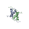
| |||||||||
|---|---|---|---|---|---|---|---|---|---|---|
| 1 |
| |||||||||
| NMR アンサンブル |
|
-要素
| #1: タンパク質・ペプチド | 分子量: 5398.181 Da / 分子数: 2 / 断片: N-terminal docking and dimerization domain / 由来タイプ: 組換発現 / 由来: (組換発現)  |
|---|
-実験情報
-実験
| 実験 | 手法: 溶液NMR |
|---|---|
| NMR実験の詳細 | Text: This structure was determined using standard homonuclear, heteronuclear, and triple resonance spectroscopy, and 3D 13C-edited(w2)-12C-filtered(w1)/13C-filtered(w3) NOESY. |
- 試料調製
試料調製
| 詳細 |
| |||||||||||||||||||||||||
|---|---|---|---|---|---|---|---|---|---|---|---|---|---|---|---|---|---|---|---|---|---|---|---|---|---|---|
| 試料状態 |
| |||||||||||||||||||||||||
| 結晶化 | *PLUS 手法: other / 詳細: NMR |
-NMR測定
| 放射 | プロトコル: SINGLE WAVELENGTH / 単色(M)・ラウエ(L): M | |||||||||||||||
|---|---|---|---|---|---|---|---|---|---|---|---|---|---|---|---|---|
| 放射波長 | 相対比: 1 | |||||||||||||||
| NMRスペクトロメーター |
|
- 解析
解析
| NMR software |
| ||||||||||||
|---|---|---|---|---|---|---|---|---|---|---|---|---|---|
| 精密化 | 手法: Hybrid distance geometry-dynamical simulated annealing, refinement protocol for monomer structure determination, with 457 NOE-derived distance restraints (185 intra-residue, i-j=0; 136 ...手法: Hybrid distance geometry-dynamical simulated annealing, refinement protocol for monomer structure determination, with 457 NOE-derived distance restraints (185 intra-residue, i-j=0; 136 sequential, |i-j|=1; 95 medium range, 1 ソフトェア番号: 1 詳細: Filtered NOESY spectrum on a 50% unlabeled-50% 13C-15N-labeled sample was used to obtain inter-molecular NOE contacts of the homodimer. Other NOEs were classified as intra-molecular and ambiguous. | ||||||||||||
| 代表構造 | 選択基準: lowest energy | ||||||||||||
| NMRアンサンブル | コンフォーマー選択の基準: structures with the least restraint violations,structures with the lowest energy 計算したコンフォーマーの数: 49 / 登録したコンフォーマーの数: 24 |
 X-PLOR