 Movie
Movie Controller

[English] 日本語
 Yorodumi
- PDB-1l6e: Solution structure of the docking and dimerization domain of prot... -
+ Open data

- Basic information
| Entry | Database: PDB / ID: 1l6e | ||||||
|---|---|---|---|---|---|---|---|
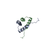
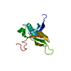
| Title | Solution structure of the docking and dimerization domain of protein kinase A II-alpha (RIIalpha D/D). Alternatively called the N-terminal dimerization domain of the regulatory subunit of protein kinase A. | ||||||
 Components Components | cAMP-dependent protein kinase Type II-alpha regulatory chain | ||||||
 Keywords Keywords | TRANSFERASE / Four-helix bundle / helix-loop-helix / regulatory subunit / dimerization / docking / anchoring | ||||||
| Function / homology |  Function and homology information Function and homology informationPKA activation in glucagon signalling / CREB1 phosphorylation through the activation of Adenylate Cyclase / DARPP-32 events / PKA activation / Vasopressin regulates renal water homeostasis via Aquaporins / GPER1 signaling / Hedgehog 'off' state / Factors involved in megakaryocyte development and platelet production / cAMP-dependent protein kinase regulator activity / High laminar flow shear stress activates signaling by PIEZO1 and PECAM1:CDH5:KDR in endothelial cells ...PKA activation in glucagon signalling / CREB1 phosphorylation through the activation of Adenylate Cyclase / DARPP-32 events / PKA activation / Vasopressin regulates renal water homeostasis via Aquaporins / GPER1 signaling / Hedgehog 'off' state / Factors involved in megakaryocyte development and platelet production / cAMP-dependent protein kinase regulator activity / High laminar flow shear stress activates signaling by PIEZO1 and PECAM1:CDH5:KDR in endothelial cells / nucleotide-activated protein kinase complex / cAMP-dependent protein kinase inhibitor activity / beta-2 adrenergic receptor binding / cAMP-dependent protein kinase complex / protein kinase A catalytic subunit binding / small molecule binding / plasma membrane raft / cAMP binding / negative regulation of cAMP/PKA signal transduction / T-tubule / modulation of chemical synaptic transmission / adenylate cyclase-activating G protein-coupled receptor signaling pathway / protein domain specific binding / ubiquitin protein ligase binding / centrosome / synapse / protein-containing complex binding / perinuclear region of cytoplasm / glutamatergic synapse / protein-containing complex / identical protein binding / plasma membrane / cytoplasm / cytosol Similarity search - Function | ||||||
| Biological species |  | ||||||
| Method | SOLUTION NMR / Hybrid distance geometry-dynamical simulated annealing, refinement protocol for monomer structure determination, with 457 NOE-derived distance restraints (185 intra-residue, i-j=0; 136 sequential, |i-j|=1; 95 medium range, 1<|i-j|<5; 41 long range, |i-j|>4), 19 distance restraints representing hydrogen bonds (entered as 2 distances each), 25 phi-, 5 chi1-torsion angle restraints. Molecular dynamical simulated annealing protocol for dimer structure determination, using 505 NOE-derived distance restraints (185 intra-residue, i-j=0; 136 sequential, |i-j|=1; 95 medium range, 1<|i-j|<5; 25 long range, |i-j|>4; 38 inter-molecular; 26 ambiguous), 19 distance restraints representing hydrogen bonds (entered as 2 distances each), 25 phi-, 5 chi1-torsion angle restraints. | ||||||
 Authors Authors | Morikis, D. / Roy, M. / Newlon, M.G. / Scott, J.D. / Jennings, P.A. | ||||||
 Citation Citation | Journal: Eur.J.Biochem. / Year: 2002 Title: Electrostatic properties of the structure of the docking and dimerization domain of protein kinase A IIalpha Authors: Morikis, D. / Roy, M. / Newlon, M.G. / Scott, J.D. / Jennings, P.A. #1: Journal: Nat.Struct.Biol. / Year: 1999Title: The molecular basis for protein kinase A anchoring revealed by solution NMR. Authors: Newlon, M.G. / Roy, M. / Morikis, D. / Hausken, Z.E. / Coghlan, V. / Scott, J.D. / Jennings, P.A. #2: Journal: J.Biol.Chem. / Year: 1997Title: The A-kinase anchoring domain of type II-alpha cAMP-dependent protein kinase is highly helical. Authors: Newlon, M.G. / Roy, M. / Hausken, Z.E. / Scott, J.D. / Jennings, P.A. | ||||||
| History |
| ||||||
| Remark 999 | SEQUENCE THE FIRST 3 RESIDUES ARE DIFFERENT DUE TO RECOMBINANT EXPRESSION AND PROTEOLYTIC CLEAVAGE. |
- Structure visualization
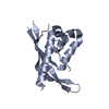
| Structure viewer | Molecule: MolmilJmol/JSmol |
|---|
- Downloads & links
-Download
| PDBx/mmCIF format | 1l6e.cif.gz | 709.4 KB | Display | PDBx/mmCIF format |
|---|---|---|---|---|
| PDB format | pdb1l6e.ent.gz | 595.7 KB | Display | PDB format |
| PDBx/mmJSON format | 1l6e.json.gz | Tree view | PDBx/mmJSON format | |
| Others |  Other downloads Other downloads |
-Validation report
| Arichive directory | https://data.pdbj.org/pub/pdb/validation_reports/l6/1l6eftp://data.pdbj.org/pub/pdb/validation_reports/l6/1l6e | HTTPS FTP |
|---|
-Related structure data
| Related structure data | |
|---|---|
| Similar structure data |


-Links
 PDBj
PDBj


- Assembly
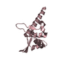
| Deposited unit | 
| |||||||||
|---|---|---|---|---|---|---|---|---|---|---|
| 1 |
| |||||||||
| NMR ensembles |
|
-Components
| #1: Protein/peptide | Mass: 5398.181 Da / Num. of mol.: 2 / Fragment: N-terminal docking and dimerization domain Source method: isolated from a genetically manipulated source Source: (gene. exp.)  |
|---|
-Experimental details
-Experiment
| Experiment | Method: SOLUTION NMR |
|---|---|
| NMR details | Text: This structure was determined using standard homonuclear, heteronuclear, and triple resonance spectroscopy, and 3D 13C-edited(w2)-12C-filtered(w1)/13C-filtered(w3) NOESY. |
- Sample preparation
| Details |
| |||||||||||||||||||||||||
|---|---|---|---|---|---|---|---|---|---|---|---|---|---|---|---|---|---|---|---|---|---|---|---|---|---|---|
| Sample conditions |
| |||||||||||||||||||||||||
| Crystal grow | *PLUS Method: other / Details: NMR |
-NMR measurement
| Radiation | Protocol: SINGLE WAVELENGTH / Monochromatic (M) / Laue (L): M | |||||||||||||||
|---|---|---|---|---|---|---|---|---|---|---|---|---|---|---|---|---|
| Radiation wavelength | Relative weight: 1 | |||||||||||||||
| NMR spectrometer |
|
- Processing
| NMR software |
| ||||||||||||
|---|---|---|---|---|---|---|---|---|---|---|---|---|---|
| Refinement | Method: Hybrid distance geometry-dynamical simulated annealing, refinement protocol for monomer structure determination, with 457 NOE-derived distance restraints (185 intra-residue, i-j=0; 136 ...Method: Hybrid distance geometry-dynamical simulated annealing, refinement protocol for monomer structure determination, with 457 NOE-derived distance restraints (185 intra-residue, i-j=0; 136 sequential, |i-j|=1; 95 medium range, 1 Software ordinal: 1 Details: Filtered NOESY spectrum on a 50% unlabeled-50% 13C-15N-labeled sample was used to obtain inter-molecular NOE contacts of the homodimer. Other NOEs were classified as intra-molecular and ambiguous. | ||||||||||||
| NMR representative | Selection criteria: lowest energy | ||||||||||||
| NMR ensemble | Conformer selection criteria: structures with the least restraint violations,structures with the lowest energy Conformers calculated total number: 49 / Conformers submitted total number: 24 |
 X-PLOR