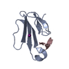
 Movie
Movie Controller


+ Open data

- Basic information
| Entry | Database: PDB / ID: 1l3d | ||||||
|---|---|---|---|---|---|---|---|
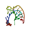
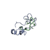
| Title | Low Resolution Crystal Structure of a Viral RNA Pseudoknot | ||||||
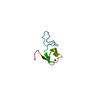
 Components Components | RNA pseudoknot | ||||||
 Keywords Keywords | RNA / Pseudoknot / frameshifting / viral RNA / flexibility | ||||||
| Function / homology | RNA / RNA (> 10) Function and homology information Function and homology information | ||||||
| Method |  X-RAY DIFFRACTION / SYNCHROTRON / MOLECULAR REPLACEMENT / Resolution: 2.85 Å X-RAY DIFFRACTION / SYNCHROTRON / MOLECULAR REPLACEMENT / Resolution: 2.85 Å | ||||||
 Authors Authors | Egli, M. / Minasov, G. / Su, L. / Rich, A. | ||||||
 Citation Citation | Journal: Proc.Natl.Acad.Sci.USA / Year: 2002 Title: Metal ions and flexibility in a viral RNA pseudoknot at atomic resolution. Authors: Egli, M. / Minasov, G. / Su, L. / Rich, A. | ||||||
| History |
|
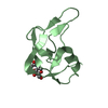
- Structure visualization
| Structure viewer | Molecule: MolmilJmol/JSmol |
|---|
- Downloads & links
-Download
| PDBx/mmCIF format | 1l3d.cif.gz | 25.8 KB | Display | PDBx/mmCIF format |
|---|---|---|---|---|
| PDB format | pdb1l3d.ent.gz | 18.1 KB | Display | PDB format |
| PDBx/mmJSON format | 1l3d.json.gz | Tree view | PDBx/mmJSON format | |
| Others |  Other downloads Other downloads |
-Validation report
| Summary document | 1l3d_validation.pdf.gz | 380.7 KB | Display | wwPDB validaton report |
|---|---|---|---|---|
| Full document | 1l3d_full_validation.pdf.gz | 382.7 KB | Display | |
| Data in XML | 1l3d_validation.xml.gz | 3.1 KB | Display | |
| Data in CIF | 1l3d_validation.cif.gz | 3.7 KB | Display | |
| Arichive directory | https://data.pdbj.org/pub/pdb/validation_reports/l3/1l3dftp://data.pdbj.org/pub/pdb/validation_reports/l3/1l3d | HTTPS FTP |
-Related structure data
| Related structure data | 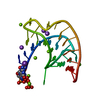 1l2xC  437dS S: Starting model for refinement C: citing same article ( |
|---|---|
| Similar structure data |


-Links
 PDBj
PDBj


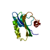
- Assembly
| Deposited unit | 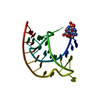
| ||||||||
|---|---|---|---|---|---|---|---|---|---|
| 1 |
| ||||||||
| Unit cell |
|
-Components
| #1: RNA chain | Mass: 9245.484 Da / Num. of mol.: 1 / Source method: obtained synthetically / Details: RNA SEQUENCE TAKEN FROM BEET WESTERN YELLOW VIRUS |
|---|---|
| #2: Water | ChemComp-HOH /  Mass: 18.015 Da / Num. of mol.: 14 / Source method: isolated from a natural source / Formula: H2O Mass: 18.015 Da / Num. of mol.: 14 / Source method: isolated from a natural source / Formula: H2O |
-Experimental details
-Experiment
| Experiment | Method: X-RAY DIFFRACTION / Number of used crystals: 1 |
|---|
- Sample preparation
| Crystal | Density Matthews: 5.65 Å3/Da / Density % sol: 78.22 % | ||||||||||||||||||||||||||||||
|---|---|---|---|---|---|---|---|---|---|---|---|---|---|---|---|---|---|---|---|---|---|---|---|---|---|---|---|---|---|---|---|
| Crystal grow | Temperature: 277 K / Method: vapor diffusion, sitting drop / pH: 5 Details: ammonium sulfate, sodium citrate, pH 5.0, VAPOR DIFFUSION, SITTING DROP, temperature 277.0K | ||||||||||||||||||||||||||||||
| Components of the solutions |
| ||||||||||||||||||||||||||||||
| Crystal grow | *PLUS Temperature: 4 ℃ | ||||||||||||||||||||||||||||||
| Components of the solutions | *PLUS
|
-Data collection
| Diffraction | Mean temperature: 100 K |
|---|---|
| Diffraction source | Source: SYNCHROTRON / Site: SSRL  / Beamline: BL1-5 / Wavelength: 1.08 Å / Beamline: BL1-5 / Wavelength: 1.08 Å |
| Detector | Type: MARRESEARCH / Detector: IMAGE PLATE / Date: Jun 17, 1997 / Details: mirrors |
| Radiation | Monochromator: graphite / Protocol: SINGLE WAVELENGTH / Monochromatic (M) / Laue (L): M / Scattering type: x-ray |
| Radiation wavelength | Wavelength: 1.08 Å / Relative weight: 1 |
| Reflection | Resolution: 2.85→30 Å / Num. all: 5034 / Num. obs: 5034 / % possible obs: 100 % / Observed criterion σ(I): -3 / Redundancy: 9.2 % / Biso Wilson estimate: 63.1 Å2 / Rmerge(I) obs: 0.054 / Net I/σ(I): 39.5 |
| Reflection shell | Resolution: 2.85→2.95 Å / Redundancy: 8.9 % / Rmerge(I) obs: 0.286 / Mean I/σ(I) obs: 9.3 / Num. unique all: 486 / % possible all: 100 |
| Reflection | *PLUS % possible obs: 100 % |
- Processing
| Software |
| |||||||||||||||||||||||||
|---|---|---|---|---|---|---|---|---|---|---|---|---|---|---|---|---|---|---|---|---|---|---|---|---|---|---|
| Refinement | Method to determine structure: MOLECULAR REPLACEMENT Starting model: PDB entry 437D Resolution: 2.85→20 Å / Isotropic thermal model: Isotropic / Cross valid method: THROUGHOUT / σ(F): 0 Stereochemistry target values: G. Parkinson et al., Acta Cryst. (1996) D52, 57-64 Details: Crystallographic conjugate gradient minimization refinement using maximum likelihood target for amplitudes
| |||||||||||||||||||||||||
| Displacement parameters | Biso mean: 43.3 Å2
| |||||||||||||||||||||||||
| Refinement step | Cycle: LAST / Resolution: 2.85→20 Å
| |||||||||||||||||||||||||
| Refine LS restraints |
| |||||||||||||||||||||||||
| LS refinement shell | Resolution: 2.85→2.98 Å
| |||||||||||||||||||||||||
| Refinement | *PLUS % reflection Rfree: 10 % / Rfactor obs: 0.271 | |||||||||||||||||||||||||
| Solvent computation | *PLUS | |||||||||||||||||||||||||
| Displacement parameters | *PLUS | |||||||||||||||||||||||||
| Refine LS restraints | *PLUS Type: c_angle_deg / Dev ideal: 1 | |||||||||||||||||||||||||
| LS refinement shell | *PLUS Rfactor obs: 0.427 |
