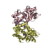
BIOMOLECULE: 1 THIS ENTRY CONTAINS THE CRYSTALLOGRAPHIC ASYMMETRIC UNIT WHICH CONSISTS OF 2 CHAIN(S) ...BIOMOLECULE: 1 THIS ENTRY CONTAINS THE CRYSTALLOGRAPHIC ASYMMETRIC UNIT WHICH CONSISTS OF 2 CHAIN(S). SEE REMARK 350 FOR INFORMATION ON GENERATING THE BIOLOGICAL MOLECULE(S). IT APPEARS THIS PROTEIN FORMS A DIMER, ALTHOUGH THERE IS NO HARD EVIDENCE EXCEPT FOR THE SIGNIFICANT INTERFACE.
Resolution: 1.7→1.76 Å / Redundancy: 2.1 % / Rmerge(I) obs: 0.244 / Mean I/σ(I) obs: 2.6 / Num. unique all: 4407 / % possible all: 81.6
-
Processing
Software
Name
Version
Classification
d*TREK
datascaling
HKL-2000
datacollection
SCALEPACK
datascaling
CNS
refinement
CrystalClear
D*STREK (MSC/RIGAKU)
datareduction
HKL-2000
datareduction
CNS
phasing
Refinement
Method to determine structure: MAD / Resolution: 1.7→43.14 Å / Rfactor Rfree error: 0.003 / Data cutoff high absF: 331388.1 / Data cutoff low absF: 0 / Isotropic thermal model: RESTRAINED / Cross valid method: THROUGHOUT / σ(F): 0 / Stereochemistry target values: Engh & Huber Details: The GLY and SER of the cloning artifact are disordered.
In the structure databanks used in Yorodumi, some data are registered as the other names, "COVID-19 virus" and "2019-nCoV". Here are the details of the virus and the list of structure data.
Jan 31, 2019. EMDB accession codes are about to change! (news from PDBe EMDB page)
EMDB accession codes are about to change! (news from PDBe EMDB page)
The allocation of 4 digits for EMDB accession codes will soon come to an end. Whilst these codes will remain in use, new EMDB accession codes will include an additional digit and will expand incrementally as the available range of codes is exhausted. The current 4-digit format prefixed with “EMD-” (i.e. EMD-XXXX) will advance to a 5-digit format (i.e. EMD-XXXXX), and so on. It is currently estimated that the 4-digit codes will be depleted around Spring 2019, at which point the 5-digit format will come into force.
The EM Navigator/Yorodumi systems omit the EMD- prefix.
Related info.:Q: What is EMD? / ID/Accession-code notation in Yorodumi/EM Navigator
Yorodumi is a browser for structure data from EMDB, PDB, SASBDB, etc.
This page is also the successor to EM Navigator detail page, and also detail information page/front-end page for Omokage search.
The word "yorodu" (or yorozu) is an old Japanese word meaning "ten thousand". "mi" (miru) is to see.
Related info.:EMDB / PDB / SASBDB / Comparison of 3 databanks / Yorodumi Search / Aug 31, 2016. New EM Navigator & Yorodumi / Yorodumi Papers / Jmol/JSmol / Function and homology information / Changes in new EM Navigator and Yorodumi
 Movie
Movie Controller


 Open data
 Basic information
 Components
 Keywords
 Function and homology information
 Thermoplasma acidophilum (acidophilic)
 X-RAY DIFFRACTION /
  Authors
 Citation
 Structure visualization
 Downloads & links
 Other downloads
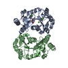


 PDBj
PDBj

 Assembly


 Mass: 40.078 Da / Num. of mol.: 6 / Source method: obtained synthetically / Formula: Ca
 Mass: 18.015 Da / Num. of mol.: 437 / Source method: isolated from a natural source / Formula: H2O
 Sample preparation
 Processing