- PDB-1ku9: X-ray Structure of a Methanococcus jannaschii DNA-Binding Protein... -
+
Open data
ID or keywords:
Loading...
-
Basic information
Entry
Database: PDB / ID: 1ku9
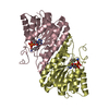
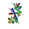
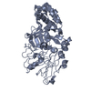
Title
X-ray Structure of a Methanococcus jannaschii DNA-Binding Protein: Implications for Antibiotic Resistance in Staphylococcus aureus
Components
hypothetical protein MJ223
Keywords
DNA BINDING PROTEIN / putative transcription factor / homodimeric winged-helix fold / STRUCTURAL GENOMICS / PSI / Protein Structure Initiative / New York SGX Research Center for Structural Genomics / NYSGXRC
Resolution: 2.8→2.9 Å / Redundancy: 5 % / Rmerge(I) obs: 0.208 / Mean I/σ(I) obs: 7.7 / Num. unique all: 941 / Rsym value: 0.208 / % possible all: 99.4
Reflection
*PLUS
Lowest resolution: 30 Å / Num. obs: 9469 / % possible obs: 99.7 % / Num. measured all: 137127
Reflection shell
*PLUS
% possible obs: 99.6 % / Rmerge(I) obs: 0.211 / Mean I/σ(I) obs: 6.9
-
Processing
Software
Name
Version
Classification
MARMAD
datacollection
DENZO
datareduction
SCALEPACK
datascaling
SnB
phasing
MLPHARE
phasing
DM
modelbuilding
CNS
1
refinement
MARMAD
datareduction
DM
phasing
Refinement
Method to determine structure: MAD / Resolution: 2.8→20 Å / Cross valid method: THROUGHOUT / σ(F): 0 / σ(I): 0 / Stereochemistry target values: Engh & Huber Details: Data were twinned and appeared to have P6(5)22 symmetry. Refined as space group P6(5) with twinning fraction of 0.485.
In the structure databanks used in Yorodumi, some data are registered as the other names, "COVID-19 virus" and "2019-nCoV". Here are the details of the virus and the list of structure data.
Jan 31, 2019. EMDB accession codes are about to change! (news from PDBe EMDB page)
EMDB accession codes are about to change! (news from PDBe EMDB page)
The allocation of 4 digits for EMDB accession codes will soon come to an end. Whilst these codes will remain in use, new EMDB accession codes will include an additional digit and will expand incrementally as the available range of codes is exhausted. The current 4-digit format prefixed with “EMD-” (i.e. EMD-XXXX) will advance to a 5-digit format (i.e. EMD-XXXXX), and so on. It is currently estimated that the 4-digit codes will be depleted around Spring 2019, at which point the 5-digit format will come into force.
The EM Navigator/Yorodumi systems omit the EMD- prefix.
Related info.:Q: What is EMD? / ID/Accession-code notation in Yorodumi/EM Navigator
Yorodumi is a browser for structure data from EMDB, PDB, SASBDB, etc.
This page is also the successor to EM Navigator detail page, and also detail information page/front-end page for Omokage search.
The word "yorodu" (or yorozu) is an old Japanese word meaning "ten thousand". "mi" (miru) is to see.
Related info.:EMDB / PDB / SASBDB / Comparison of 3 databanks / Yorodumi Search / Aug 31, 2016. New EM Navigator & Yorodumi / Yorodumi Papers / Jmol/JSmol / Function and homology information / Changes in new EM Navigator and Yorodumi
 Movie
Movie Controller

 Yorodumi
 Open data
 Basic information
 Components
 Keywords
 Function and homology information
 Methanocaldococcus jannaschii (archaea)
 X-RAY DIFFRACTION /
  Authors
 Citation
 Structure visualization
 Downloads & links
 Other downloads
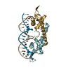


 PDBj
PDBj Assembly
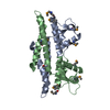

 Mass: 18.015 Da / Num. of mol.: 86 / Source method: isolated from a natural source / Formula: H2O
 Sample preparation
 / Beamline: X12C / Wavelength: 0.9793, 0.9790, 0.9686
 Processing