 Movie
Movie Controller


+ Open data

- Basic information
| Entry | Database: PDB / ID: 1kt6 | ||||||
|---|---|---|---|---|---|---|---|
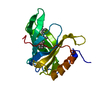
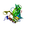
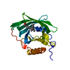
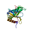
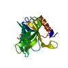
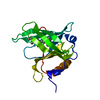
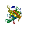
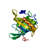
| Title | Crystal structure of bovine holo-RBP at pH 9.0 | ||||||
 Components Components | plasma retinol-binding protein | ||||||
 Keywords Keywords | TRANSPORT PROTEIN / RBP / retinol binding | ||||||
| Function / homology |  Function and homology information Function and homology informationretinol transport / retinol transmembrane transporter activity / retinal binding / retinol binding / : Similarity search - Function | ||||||
| Biological species |  | ||||||
| Method |  X-RAY DIFFRACTION / SYNCHROTRON / MOLECULAR REPLACEMENT / Resolution: 1.1 Å X-RAY DIFFRACTION / SYNCHROTRON / MOLECULAR REPLACEMENT / Resolution: 1.1 Å | ||||||
 Authors Authors | Calderone, V. / Berni, R. / Zanotti, G. | ||||||
 Citation Citation | Journal: J.Mol.Biol. / Year: 2003 Title: High-resolution Structures of Retinol-binding Protein in Complex with Retinol: pH-induced Protein Structural Changes in the Crystal State Authors: Calderone, V. / Berni, R. / Zanotti, G. #1: Journal: Proteins / Year: 1990Title: Crystallographic Refinement of Human Serum Retinol Binding Protein at 2A Resolution. Authors: Cowan, S.W. / Newcomer, M.E. / Jones, T.A. #2: Journal: J.Biol.Chem. / Year: 1993Title: Crystal Structure of Liganded and Unliganded Forms of Bovine Plasma Retinol-Binding Protein Authors: Zanotti, G. / Berni, R. / Monaco, H.L. | ||||||
| History |
|
- Structure visualization
| Structure viewer | Molecule: MolmilJmol/JSmol |
|---|
- Downloads & links
-Download
| PDBx/mmCIF format | 1kt6.cif.gz | 92.2 KB | Display | PDBx/mmCIF format |
|---|---|---|---|---|
| PDB format | pdb1kt6.ent.gz | 70 KB | Display | PDB format |
| PDBx/mmJSON format | 1kt6.json.gz | Tree view | PDBx/mmJSON format | |
| Others |  Other downloads Other downloads |
-Validation report
| Arichive directory | https://data.pdbj.org/pub/pdb/validation_reports/kt/1kt6ftp://data.pdbj.org/pub/pdb/validation_reports/kt/1kt6 | HTTPS FTP |
|---|
-Related structure data
| Related structure data |  1kt3C  1kt4C  1kt5C 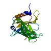 1kt7C  1hbpS S: Starting model for refinement C: citing same article ( |
|---|---|
| Similar structure data |


-Links
 PDBj
PDBj

- Assembly
| Deposited unit | 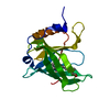
| ||||||||
|---|---|---|---|---|---|---|---|---|---|
| 1 |
| ||||||||
| Unit cell |
|
-Components
| #1: Protein | Mass: 21095.654 Da / Num. of mol.: 1 / Source method: isolated from a natural source / Source: (natural) |
|---|---|
| #2: Chemical | ChemComp-RTL /   Mass: 286.452 Da / Num. of mol.: 1 / Source method: obtained synthetically / Formula: C20H30O Mass: 286.452 Da / Num. of mol.: 1 / Source method: obtained synthetically / Formula: C20H30O |
| #3: Water | ChemComp-HOH /  Mass: 18.015 Da / Num. of mol.: 172 / Source method: isolated from a natural source / Formula: H2O Mass: 18.015 Da / Num. of mol.: 172 / Source method: isolated from a natural source / Formula: H2O |
| Has protein modification | Y |
-Experimental details
-Experiment
| Experiment | Method: X-RAY DIFFRACTION / Number of used crystals: 1 |
|---|
- Sample preparation
| Crystal | Density Matthews: 1.82 Å3/Da / Density % sol: 40.17 % | |||||||||||||||||||||||||||||||||||
|---|---|---|---|---|---|---|---|---|---|---|---|---|---|---|---|---|---|---|---|---|---|---|---|---|---|---|---|---|---|---|---|---|---|---|---|---|
| Crystal grow | Temperature: 293 K / Method: vapor diffusion, sitting drop / pH: 9 Details: 100 mM HEPES, 50 mM NaCl, pH 9.0, VAPOR DIFFUSION, SITTING DROP, temperature 293K | |||||||||||||||||||||||||||||||||||
| Crystal grow | *PLUS Temperature: 20 ℃ / pH: 7 / Method: vapor diffusion / Details: used microseeding | |||||||||||||||||||||||||||||||||||
| Components of the solutions | *PLUS
|
-Data collection
| Diffraction | Mean temperature: 100 K |
|---|---|
| Diffraction source | Source: SYNCHROTRON / Site: ESRF  / Beamline: ID14-4 / Wavelength: 0.9393 Å / Beamline: ID14-4 / Wavelength: 0.9393 Å |
| Detector | Type: MARRESEARCH / Detector: CCD / Date: Feb 10, 2001 |
| Radiation | Protocol: SINGLE WAVELENGTH / Monochromatic (M) / Laue (L): M / Scattering type: x-ray |
| Radiation wavelength | Wavelength: 0.9393 Å / Relative weight: 1 |
| Reflection | Resolution: 1.098→22.418 Å / Num. all: 62345 / Num. obs: 62345 / % possible obs: 87.7 % / Observed criterion σ(F): 0 / Observed criterion σ(I): 0 / Redundancy: 3.6 % / Rmerge(I) obs: 0.066 / Rsym value: 0.066 / Net I/σ(I): 5.7 |
| Reflection shell | Resolution: 1.1→1.16 Å / Redundancy: 2.6 % / Rmerge(I) obs: 0.228 / Mean I/σ(I) obs: 2.4 / Num. unique all: 5808 / Rsym value: 0.228 / % possible all: 70.3 |
| Reflection | *PLUS Highest resolution: 1.09 Å / Lowest resolution: 40.82 Å / Num. obs: 65032 / % possible obs: 88.2 % / Redundancy: 3.8 % / Num. measured all: 552856 / Rmerge(I) obs: 0.067 |
| Reflection shell | *PLUS % possible obs: 70.3 % |
- Processing
| Software |
| |||||||||||||||||||||||||||||||||
|---|---|---|---|---|---|---|---|---|---|---|---|---|---|---|---|---|---|---|---|---|---|---|---|---|---|---|---|---|---|---|---|---|---|---|
| Refinement | Method to determine structure: MOLECULAR REPLACEMENT Starting model: 1HBP Resolution: 1.1→22.418 Å / Num. parameters: 14538 / Num. restraintsaints: 17752 / Isotropic thermal model: anisotropic / Cross valid method: FREE R / σ(F): 0 / Stereochemistry target values: ENGH AND HUBER
| |||||||||||||||||||||||||||||||||
| Solvent computation | Solvent model: MOEWS & KRETSINGER, J.MOL.BIOL.91(1973)201-228 | |||||||||||||||||||||||||||||||||
| Refine analyze | Num. disordered residues: 0 / Occupancy sum hydrogen: 1330 / Occupancy sum non hydrogen: 1615 | |||||||||||||||||||||||||||||||||
| Refinement step | Cycle: LAST / Resolution: 1.1→22.418 Å
| |||||||||||||||||||||||||||||||||
| Refine LS restraints |
| |||||||||||||||||||||||||||||||||
| Software | *PLUS Name: SHELXL / Version: 97 / Classification: refinement | |||||||||||||||||||||||||||||||||
| Refinement | *PLUS Num. reflection obs: 65032 / Num. reflection Rfree: 3252 | |||||||||||||||||||||||||||||||||
| Solvent computation | *PLUS | |||||||||||||||||||||||||||||||||
| Displacement parameters | *PLUS | |||||||||||||||||||||||||||||||||
| Refine LS restraints | *PLUS
|
