 Movie
Movie Controller

[English] 日本語
 Yorodumi
- PDB-1ksc: The structure of Endoglucanase from termite, Nasutitermes takasag... -
+ Open data

- Basic information
| Entry | Database: PDB / ID: 1ksc | ||||||
|---|---|---|---|---|---|---|---|
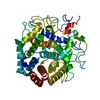
| Title | The structure of Endoglucanase from termite, Nasutitermes takasagoensis, at pH 5.6. | ||||||
 Components Components | Endo-b-1,4-glucanase | ||||||
 Keywords Keywords | HYDROLASE / Cellulase / Endoglucanase / Termite / Nasutitermes takasagoensis / Glycosyl Hydrolase / Family 9 / (Alpha/Alpha)6 | ||||||
| Function / homology |  Function and homology information Function and homology informationcellulase / cellulase activity / cellulose catabolic process / metal ion binding Similarity search - Function | ||||||
| Biological species |  Nasutitermes takasagoensis (cockroach) Nasutitermes takasagoensis (cockroach) | ||||||
| Method |  X-RAY DIFFRACTION / MOLECULAR REPLACEMENT / Resolution: 1.55 Å X-RAY DIFFRACTION / MOLECULAR REPLACEMENT / Resolution: 1.55 Å | ||||||
 Authors Authors | Khademi, S. / Guarino, L.A. / Watanabe, H. / Tokuda, G. / Meyer, E.F. | ||||||
 Citation Citation | Journal: Acta Crystallogr.,Sect.D / Year: 2002 Title: Structure of an endoglucanase from termite, Nasutitermes takasagoensis. Authors: Khademi, S. / Guarino, L.A. / Watanabe, H. / Tokuda, G. / Meyer, E.F. | ||||||
| History |
|
- Structure visualization
| Structure viewer | Molecule: MolmilJmol/JSmol |
|---|
- Downloads & links
-Download
| PDBx/mmCIF format | 1ksc.cif.gz | 110.1 KB | Display | PDBx/mmCIF format |
|---|---|---|---|---|
| PDB format | pdb1ksc.ent.gz | 82.9 KB | Display | PDB format |
| PDBx/mmJSON format | 1ksc.json.gz | Tree view | PDBx/mmJSON format | |
| Others |  Other downloads Other downloads |
-Validation report
| Summary document | 1ksc_validation.pdf.gz | 413.3 KB | Display | wwPDB validaton report |
|---|---|---|---|---|
| Full document | 1ksc_full_validation.pdf.gz | 417.1 KB | Display | |
| Data in XML | 1ksc_validation.xml.gz | 22.6 KB | Display | |
| Data in CIF | 1ksc_validation.cif.gz | 35.4 KB | Display | |
| Arichive directory | https://data.pdbj.org/pub/pdb/validation_reports/ks/1kscftp://data.pdbj.org/pub/pdb/validation_reports/ks/1ksc | HTTPS FTP |
-Related structure data
| Related structure data |  1ks8C 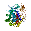 1ksdC  3tf4S C: citing same article ( S: Starting model for refinement |
|---|---|
| Similar structure data |


-Links
 PDBj
PDBj
- Assembly
| Deposited unit | 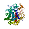
| ||||||||
|---|---|---|---|---|---|---|---|---|---|
| 1 |
| ||||||||
| Unit cell |
|
-Components
| #1: Protein | Mass: 47844.309 Da / Num. of mol.: 1 / Fragment: Catalytic Domain Source method: isolated from a genetically manipulated source Source: (gene. exp.) Nasutitermes takasagoensis (cockroach) / Plasmid: pET3a-Nts / Species (production host): Escherichia coli / Production host:  |
|---|---|
| #2: Chemical | ChemComp-CA /   Mass: 40.078 Da / Num. of mol.: 1 / Source method: obtained synthetically / Formula: Ca Mass: 40.078 Da / Num. of mol.: 1 / Source method: obtained synthetically / Formula: Ca |
| #3: Water | ChemComp-HOH /  Mass: 18.015 Da / Num. of mol.: 567 / Source method: isolated from a natural source / Formula: H2O Mass: 18.015 Da / Num. of mol.: 567 / Source method: isolated from a natural source / Formula: H2O |
| Has protein modification | Y |
-Experimental details
-Experiment
| Experiment | Method: X-RAY DIFFRACTION / Number of used crystals: 1 |
|---|
- Sample preparation
| Crystal | Density Matthews: 2.37 Å3/Da / Density % sol: 48.01 % | ||||||||||||||||||||||||
|---|---|---|---|---|---|---|---|---|---|---|---|---|---|---|---|---|---|---|---|---|---|---|---|---|---|
| Crystal grow | Temperature: 291 K / Method: vapor diffusion, hanging drop / pH: 5.6 Details: Ammonium Sulfate, Sodium Citrate, pH 4.5, VAPOR DIFFUSION, HANGING DROP, temperature 291K, pH 5.6 | ||||||||||||||||||||||||
| Crystal grow | *PLUS pH: 4.5 | ||||||||||||||||||||||||
| Components of the solutions | *PLUS
|
-Data collection
| Diffraction | Mean temperature: 120 K |
|---|---|
| Diffraction source | Source: ROTATING ANODE / Type: RIGAKU RU200 / Wavelength: 1.5418 Å |
| Detector | Type: MAC Science DIP-2030K / Detector: IMAGE PLATE / Date: Jun 1, 2000 / Details: mirrors |
| Radiation | Monochromator: OSMIC, Model 140-000023 / Protocol: SINGLE WAVELENGTH / Monochromatic (M) / Laue (L): M / Scattering type: x-ray |
| Radiation wavelength | Wavelength: 1.5418 Å / Relative weight: 1 |
| Reflection | Resolution: 1.45→30 Å / Num. all: 61593 / Num. obs: 70043 / % possible obs: 93.3 % / Observed criterion σ(F): 1 / Observed criterion σ(I): 3 / Redundancy: 18.73 % / Biso Wilson estimate: 16.2 Å2 / Rmerge(I) obs: 0.079 / Net I/σ(I): 19.6 |
| Reflection shell | Resolution: 1.45→1.5 Å / Rmerge(I) obs: 0.079 / Mean I/σ(I) obs: 3.6 / % possible all: 83.8 |
| Reflection | *PLUS Highest resolution: 1.55 Å / Lowest resolution: 30 Å / Num. obs: 60004 / Num. measured all: 1124141 |
| Reflection shell | *PLUS Highest resolution: 1.55 Å / Lowest resolution: 1.61 Å / % possible obs: 83.8 % / Rmerge(I) obs: 0.562 |
- Processing
| Software |
| ||||||||||||||||||||||||||||||||||||
|---|---|---|---|---|---|---|---|---|---|---|---|---|---|---|---|---|---|---|---|---|---|---|---|---|---|---|---|---|---|---|---|---|---|---|---|---|---|
| Refinement | Method to determine structure: MOLECULAR REPLACEMENT Starting model: 3TF4 Resolution: 1.55→30 Å / Cross valid method: THROUGHOUT / σ(F): 0 / Stereochemistry target values: Engh & Huber
| ||||||||||||||||||||||||||||||||||||
| Solvent computation | Solvent model: 0.373825 / ksol: 56.4412 e/Å3 | ||||||||||||||||||||||||||||||||||||
| Displacement parameters | Biso mean: 18.5 Å2
| ||||||||||||||||||||||||||||||||||||
| Refine analyze |
| ||||||||||||||||||||||||||||||||||||
| Refinement step | Cycle: LAST / Resolution: 1.55→30 Å
| ||||||||||||||||||||||||||||||||||||
| Refine LS restraints |
| ||||||||||||||||||||||||||||||||||||
| LS refinement shell | Resolution: 1.55→1.61 Å / Rfactor Rfree error: 0.013 / Total num. of bins used: 10
| ||||||||||||||||||||||||||||||||||||
| Xplor file | Serial no: 1 / Param file: protein_rep.param / Topol file: protein.top | ||||||||||||||||||||||||||||||||||||
| Refinement | *PLUS Lowest resolution: 30 Å | ||||||||||||||||||||||||||||||||||||
| Solvent computation | *PLUS | ||||||||||||||||||||||||||||||||||||
| Displacement parameters | *PLUS | ||||||||||||||||||||||||||||||||||||
| Refine LS restraints | *PLUS
|
