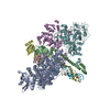
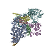
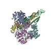
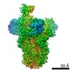
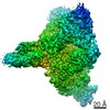
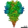
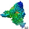
Entry Database : PDB / ID : 1kqgTitle FORMATE DEHYDROGENASE N FROM E. COLI (FORMATE DEHYDROGENASE, NITRATE-INDUCIBLE, ...) x 3 Keywords / / / / / / / / / / / / / / Function / homology Function Domain/homology Component
/ / / / / / / / / / / / / / / / / / / / / / / / / / / / / / / / / / / / / / / / / / / / / / / / / / / / / / / / / / / / / / / / / / / / / / / / / / / / / / / / / / / / / / / / / / / / / / / / / / / / Biological species Escherichia coli (E. coli)Method / / / Resolution : 2.8 Å Authors Jormakka, M. / Tornroth, S. / Byrne, B. / Iwata, S. Journal : Science / Year : 2002Title : Molecular basis of proton motive force generation: structure of formate dehydrogenase-N.Authors : Jormakka, M. / Tornroth, S. / Byrne, B. / Iwata, S. History Deposition Jan 5, 2002 Deposition site / Processing site Revision 1.0 Mar 15, 2002 Provider / Type Revision 1.1 Apr 2, 2008 Group Revision 1.2 Jul 13, 2011 Group / Version format complianceRevision 1.3 Feb 19, 2014 Group / Structure summaryRevision 1.4 Aug 6, 2014 Group / Structure summaryRevision 1.5 Feb 14, 2024 Group / Database references / Derived calculationsCategory chem_comp_atom / chem_comp_bond ... chem_comp_atom / chem_comp_bond / database_2 / pdbx_struct_conn_angle / struct_conn / struct_site Item _database_2.pdbx_DOI / _database_2.pdbx_database_accession ... _database_2.pdbx_DOI / _database_2.pdbx_database_accession / _pdbx_struct_conn_angle.ptnr1_auth_asym_id / _pdbx_struct_conn_angle.ptnr1_auth_comp_id / _pdbx_struct_conn_angle.ptnr1_auth_seq_id / _pdbx_struct_conn_angle.ptnr1_label_asym_id / _pdbx_struct_conn_angle.ptnr1_label_atom_id / _pdbx_struct_conn_angle.ptnr1_label_comp_id / _pdbx_struct_conn_angle.ptnr1_label_seq_id / _pdbx_struct_conn_angle.ptnr2_auth_asym_id / _pdbx_struct_conn_angle.ptnr2_auth_comp_id / _pdbx_struct_conn_angle.ptnr2_auth_seq_id / _pdbx_struct_conn_angle.ptnr2_label_asym_id / _pdbx_struct_conn_angle.ptnr2_label_atom_id / _pdbx_struct_conn_angle.ptnr2_label_comp_id / _pdbx_struct_conn_angle.ptnr3_auth_asym_id / _pdbx_struct_conn_angle.ptnr3_auth_comp_id / _pdbx_struct_conn_angle.ptnr3_auth_seq_id / _pdbx_struct_conn_angle.ptnr3_label_asym_id / _pdbx_struct_conn_angle.ptnr3_label_atom_id / _pdbx_struct_conn_angle.ptnr3_label_comp_id / _pdbx_struct_conn_angle.ptnr3_label_seq_id / _pdbx_struct_conn_angle.value / _struct_conn.pdbx_dist_value / _struct_conn.ptnr1_auth_asym_id / _struct_conn.ptnr1_auth_comp_id / _struct_conn.ptnr1_auth_seq_id / _struct_conn.ptnr1_label_asym_id / _struct_conn.ptnr1_label_atom_id / _struct_conn.ptnr1_label_comp_id / _struct_conn.ptnr1_label_seq_id / _struct_conn.ptnr2_auth_asym_id / _struct_conn.ptnr2_auth_comp_id / _struct_conn.ptnr2_auth_seq_id / _struct_conn.ptnr2_label_asym_id / _struct_conn.ptnr2_label_atom_id / _struct_conn.ptnr2_label_comp_id / _struct_site.pdbx_auth_asym_id / _struct_site.pdbx_auth_comp_id / _struct_site.pdbx_auth_seq_id
Show all Show less
 Movie
Movie Controller


 Open data
 Basic information
 Components
 Keywords
 Function and homology information
 X-RAY DIFFRACTION /
  Authors
 Citation
 Structure visualization
 Downloads & links
 Other downloads

 PDBj
PDBj


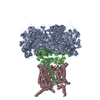
 Assembly
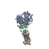


 Mass: 351.640 Da / Num. of mol.: 5 / Source method: obtained synthetically / Formula: Fe4S4
 Mass: 740.557 Da / Num. of mol.: 2 / Source method: obtained synthetically / Formula: C20H26N10O13P2S2
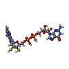
 Mass: 95.940 Da / Num. of mol.: 1 / Source method: obtained synthetically / Formula: Mo
 Mass: 1464.043 Da / Num. of mol.: 1 / Source method: obtained synthetically / Formula: C81H156O17P2 / Comment: phospholipid*YM
 Mass: 616.487 Da / Num. of mol.: 2 / Source method: obtained synthetically / Formula: C34H32FeN4O4
 Mass: 259.343 Da / Num. of mol.: 1 / Source method: obtained synthetically / Formula: C16H21NO2
 Sample preparation
 / Beamline: ID14-2 / Wavelength: 0.915 Å
 Processing