 Movie
Movie Controller


+ Open data

- Basic information
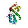
| Entry | Database: PDB / ID: 1klx | ||||||
|---|---|---|---|---|---|---|---|
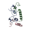
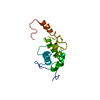
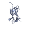
| Title | Helicobacter pylori cysteine rich protein B (hcpB) | ||||||
 Components Components | Cysteine Rich Protein B | ||||||
 Keywords Keywords | HYDROLASE / Structural Genomics / helix-turn-helix / right handed super helix / modular structure' | ||||||
| Function / homology |  Function and homology information Function and homology information | ||||||
| Biological species |   Helicobacter pylori (bacteria) Helicobacter pylori (bacteria) | ||||||
| Method |  X-RAY DIFFRACTION / SYNCHROTRON / MAD / Resolution: 1.95 Å X-RAY DIFFRACTION / SYNCHROTRON / MAD / Resolution: 1.95 Å | ||||||
 Authors Authors | Luethy, L. / Gruetter, M.G. / Mittl, P.R.E. | ||||||
 Citation Citation | Journal: J.Biol.Chem. / Year: 2002 Title: The crystal structure of Helicobacter pylori cysteine-rich protein B reveals a novel fold for a penicillin-binding protein. Authors: Luthy, L. / Grutter, M.G. / Mittl, P.R. | ||||||
| History |
|
- Structure visualization
| Structure viewer | Molecule: MolmilJmol/JSmol |
|---|
- Downloads & links
-Download
| PDBx/mmCIF format | 1klx.cif.gz | 41.6 KB | Display | PDBx/mmCIF format |
|---|---|---|---|---|
| PDB format | pdb1klx.ent.gz | 29.7 KB | Display | PDB format |
| PDBx/mmJSON format | 1klx.json.gz | Tree view | PDBx/mmJSON format | |
| Others |  Other downloads Other downloads |
-Validation report
| Arichive directory | https://data.pdbj.org/pub/pdb/validation_reports/kl/1klxftp://data.pdbj.org/pub/pdb/validation_reports/kl/1klx | HTTPS FTP |
|---|
-Related structure data
| Similar structure data |
|---|


-Links
 PDBj
PDBj- Assembly
| Deposited unit | 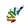
| ||||||||
|---|---|---|---|---|---|---|---|---|---|
| 1 |
| ||||||||
| Unit cell |
|
-Components
| #1: Protein | Mass: 15367.736 Da / Num. of mol.: 1 Source method: isolated from a genetically manipulated source Source: (gene. exp.) Helicobacter pylori (bacteria) / Gene: HP0336 / Plasmid: pTFT74 / Species (production host): Escherichia coli / Production host: |
|---|---|
| #2: Water | ChemComp-HOH /  Mass: 18.015 Da / Num. of mol.: 189 / Source method: isolated from a natural source / Formula: H2O Mass: 18.015 Da / Num. of mol.: 189 / Source method: isolated from a natural source / Formula: H2O |
| Has protein modification | Y |
-Experimental details
-Experiment
| Experiment | Method: X-RAY DIFFRACTION / Number of used crystals: 2 |
|---|
- Sample preparation
| Crystal | Density Matthews: 2.4 Å3/Da / Density % sol: 48.9 % | ||||||||||||||||||||||||||||||||||||
|---|---|---|---|---|---|---|---|---|---|---|---|---|---|---|---|---|---|---|---|---|---|---|---|---|---|---|---|---|---|---|---|---|---|---|---|---|---|
| Crystal grow | Temperature: 293 K / Method: vapor diffusion, sitting drop / pH: 3 Details: PEG 8000, sodium citrate, pH 3, VAPOR DIFFUSION, SITTING DROP, temperature 293K | ||||||||||||||||||||||||||||||||||||
| Crystal grow | *PLUS Temperature: 20 ℃ / pH: 5.5 | ||||||||||||||||||||||||||||||||||||
| Components of the solutions | *PLUS
|
-Data collection
| Diffraction |
| |||||||||||||||
|---|---|---|---|---|---|---|---|---|---|---|---|---|---|---|---|---|
| Diffraction source |
| |||||||||||||||
| Radiation | Protocol: SINGLE WAVELENGTH / Monochromatic (M) / Laue (L): M / Scattering type: x-ray | |||||||||||||||
| Radiation wavelength |
| |||||||||||||||
| Reflection | Resolution: 1.95→30 Å / Num. all: 12606 / Num. obs: 12049 / % possible obs: 95.6 % / Observed criterion σ(F): 2 / Observed criterion σ(I): 2 | |||||||||||||||
| Reflection shell | Resolution: 1.95→2.001 Å / % possible all: 95.6 | |||||||||||||||
| Reflection | *PLUS Lowest resolution: 30 Å / % possible obs: 99.8 % / Redundancy: 9.3 % / Rmerge(I) obs: 0.054 | |||||||||||||||
| Reflection shell | *PLUS Lowest resolution: 2 Å / % possible obs: 49.8 % |

- Processing
| Software |
| ||||||||||||||||||||||||||||||||||||||||||||||||||||||||||||||||||||||||||||||||||||||||||||||||||||||||||||||||||||||||
|---|---|---|---|---|---|---|---|---|---|---|---|---|---|---|---|---|---|---|---|---|---|---|---|---|---|---|---|---|---|---|---|---|---|---|---|---|---|---|---|---|---|---|---|---|---|---|---|---|---|---|---|---|---|---|---|---|---|---|---|---|---|---|---|---|---|---|---|---|---|---|---|---|---|---|---|---|---|---|---|---|---|---|---|---|---|---|---|---|---|---|---|---|---|---|---|---|---|---|---|---|---|---|---|---|---|---|---|---|---|---|---|---|---|---|---|---|---|---|---|---|---|
| Refinement | Method to determine structure: MAD / Resolution: 1.95→30 Å / Cor.coef. Fo:Fc: 0.951 / SU B: 3.738 / SU ML: 0.108 / Cross valid method: THROUGHOUT / σ(F): 0 / ESU R: 0.173 / ESU R Free: 0.164
| ||||||||||||||||||||||||||||||||||||||||||||||||||||||||||||||||||||||||||||||||||||||||||||||||||||||||||||||||||||||||
| Solvent computation | Solvent model: BABINET MODEL WITH MASK | ||||||||||||||||||||||||||||||||||||||||||||||||||||||||||||||||||||||||||||||||||||||||||||||||||||||||||||||||||||||||
| Displacement parameters | Biso mean: 32.926 Å2
| ||||||||||||||||||||||||||||||||||||||||||||||||||||||||||||||||||||||||||||||||||||||||||||||||||||||||||||||||||||||||
| Refinement step | Cycle: LAST / Resolution: 1.95→30 Å
| ||||||||||||||||||||||||||||||||||||||||||||||||||||||||||||||||||||||||||||||||||||||||||||||||||||||||||||||||||||||||
| Refine LS restraints |
| ||||||||||||||||||||||||||||||||||||||||||||||||||||||||||||||||||||||||||||||||||||||||||||||||||||||||||||||||||||||||
| LS refinement shell | Resolution: 1.95→2.001 Å / Total num. of bins used: 20 /
| ||||||||||||||||||||||||||||||||||||||||||||||||||||||||||||||||||||||||||||||||||||||||||||||||||||||||||||||||||||||||
| Refinement | *PLUS Highest resolution: 1.95 Å / Lowest resolution: 30 Å / Rfactor Rfree: 0.2384 / Rfactor Rwork: 0.1867 | ||||||||||||||||||||||||||||||||||||||||||||||||||||||||||||||||||||||||||||||||||||||||||||||||||||||||||||||||||||||||
| Solvent computation | *PLUS | ||||||||||||||||||||||||||||||||||||||||||||||||||||||||||||||||||||||||||||||||||||||||||||||||||||||||||||||||||||||||
| Displacement parameters | *PLUS |
