 Movie
Movie Controller


+ Open data

- Basic information
| Entry | Database: PDB / ID: 1kil | ||||||
|---|---|---|---|---|---|---|---|
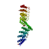
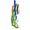
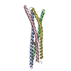
| Title | Three-dimensional structure of the complexin/SNARE complex | ||||||
 Components Components |
| ||||||
 Keywords Keywords | MEMBRANE PROTEIN / Helix bound to four helix bundle | ||||||
| Function / homology |  Function and homology information Function and homology informationregulation of exocytic insertion of neurotransmitter receptor to postsynaptic membrane / Toxicity of botulinum toxin type C (botC) / regulation of synaptic vesicle fusion to presynaptic active zone membrane / exocytic insertion of neurotransmitter receptor to postsynaptic membrane / trans-Golgi Network Vesicle Budding / regulation of delayed rectifier potassium channel activity / neurotransmitter uptake / Toxicity of botulinum toxin type E (botE) / myosin head/neck binding / Lysosome Vesicle Biogenesis ...regulation of exocytic insertion of neurotransmitter receptor to postsynaptic membrane / Toxicity of botulinum toxin type C (botC) / regulation of synaptic vesicle fusion to presynaptic active zone membrane / exocytic insertion of neurotransmitter receptor to postsynaptic membrane / trans-Golgi Network Vesicle Budding / regulation of delayed rectifier potassium channel activity / neurotransmitter uptake / Toxicity of botulinum toxin type E (botE) / myosin head/neck binding / Lysosome Vesicle Biogenesis / GABA synthesis, release, reuptake and degradation / zymogen granule membrane / synaptic vesicle fusion to presynaptic active zone membrane / storage vacuole / Other interleukin signaling / synaptobrevin 2-SNAP-25-syntaxin-1a-complexin II complex / Toxicity of botulinum toxin type A (botA) / synaptobrevin 2-SNAP-25-syntaxin-1a complex / Acetylcholine Neurotransmitter Release Cycle / synaptobrevin 2-SNAP-25-syntaxin-1a-complexin I complex / presynaptic dense core vesicle exocytosis / Glutamate Neurotransmitter Release Cycle / Norepinephrine Neurotransmitter Release Cycle / Acetylcholine Neurotransmitter Release Cycle / Serotonin Neurotransmitter Release Cycle / Serotonin Neurotransmitter Release Cycle / GABA synthesis, release, reuptake and degradation / positive regulation of catecholamine secretion / positive regulation of norepinephrine secretion / Dopamine Neurotransmitter Release Cycle / vesicle-mediated transport in synapse / eosinophil degranulation / regulation of synaptic vesicle priming / regulated exocytosis / Golgi Associated Vesicle Biogenesis / : / Insertion of tail-anchored proteins into the endoplasmic reticulum membrane / Norepinephrine Neurotransmitter Release Cycle / Dopamine Neurotransmitter Release Cycle / SNARE complex disassembly / positive regulation of calcium ion-dependent exocytosis / positive regulation of intracellular protein transport / ribbon synapse / regulation of vesicle-mediated transport / : / chloride channel inhibitor activity / secretion by cell / Glutamate Neurotransmitter Release Cycle / Cargo recognition for clathrin-mediated endocytosis / regulation of exocytosis / calcium-ion regulated exocytosis / Clathrin-mediated endocytosis / SNARE complex / SNAP receptor activity / vesicle fusion / actomyosin / LGI-ADAM interactions / hormone secretion / Golgi to plasma membrane protein transport / response to cholesterol / ATP-dependent protein binding / clathrin-coated vesicle / syntaxin binding / protein localization to membrane / syntaxin-1 binding / insulin secretion / Other interleukin signaling / Sensory processing of sound by inner hair cells of the cochlea / regulation of synaptic vesicle recycling / myosin binding / positive regulation of neurotransmitter secretion / regulation of neuron projection development / SNARE complex assembly / synaptic vesicle priming / neuron projection terminus / neurotransmitter transport / response to gravity / regulation of insulin secretion / exocytosis / positive regulation of exocytosis / tertiary granule membrane / synaptic vesicle exocytosis / associative learning / modulation of excitatory postsynaptic potential / protein sumoylation / voltage-gated potassium channel activity / postsynaptic cytosol / synaptic vesicle endocytosis / response to glucose / specific granule membrane / calcium channel inhibitor activity / vesicle-mediated transport / photoreceptor inner segment / establishment of localization in cell / somatodendritic compartment / endomembrane system / presynaptic active zone membrane / secretory granule / acrosomal vesicle / cytoplasmic vesicle membrane Similarity search - Function | ||||||
| Biological species |   Homo sapiens (human) Homo sapiens (human) | ||||||
| Method |  X-RAY DIFFRACTION / SYNCHROTRON / MOLECULAR REPLACEMENT / Resolution: 2.3 Å X-RAY DIFFRACTION / SYNCHROTRON / MOLECULAR REPLACEMENT / Resolution: 2.3 Å | ||||||
 Authors Authors | Chen, X. / Tomchick, D. / Kovrigin, E. / Arac, D. / Machius, M. / Sudhof, T.C. / Rizo, J. | ||||||
 Citation Citation | Journal: Neuron / Year: 2002 Title: Three-dimensional structure of the complexin/SNARE complex. Authors: Chen, X. / Tomchick, D.R. / Kovrigin, E. / Arac, D. / Machius, M. / Sudhof, T.C. / Rizo, J. | ||||||
| History |
| ||||||
| Remark 999 | SEQUENCE RESIDUE 10 CHAIN C, THE WILD-TYPE SEQUENCE STARTS WITH LEU11. SER10 IN THE COORDINATES IS ... SEQUENCE RESIDUE 10 CHAIN C, THE WILD-TYPE SEQUENCE STARTS WITH LEU11. SER10 IN THE COORDINATES IS PART OF THE VECTOR GLY9 WHICH IS DISORDERED. RESIDUE 140 CHAIN D, THE WILD-TYPE SEQUENCE STARTS WITH ALA141. BOTH RESIDUES FROM THE VECTOR GLY139 AND SER140 ARE ORDERED. RESIDUE 204 CHAIN D IS AN ENGINEERED TRP(FLUORESCENCE STUDIES) IT IS NOT PART OF THE NATIVE SEQUENCES, THREFORE NOT A MUTATION. |
- Structure visualization
| Structure viewer | Molecule: MolmilJmol/JSmol |
|---|
- Downloads & links
-Download
| PDBx/mmCIF format | 1kil.cif.gz | 80.2 KB | Display | PDBx/mmCIF format |
|---|---|---|---|---|
| PDB format | pdb1kil.ent.gz | 59.2 KB | Display | PDB format |
| PDBx/mmJSON format | 1kil.json.gz | Tree view | PDBx/mmJSON format | |
| Others |  Other downloads Other downloads |
-Validation report
| Arichive directory | https://data.pdbj.org/pub/pdb/validation_reports/ki/1kilftp://data.pdbj.org/pub/pdb/validation_reports/ki/1kil | HTTPS FTP |
|---|
-Related structure data
| Related structure data |  1sfcS S: Starting model for refinement |
|---|---|
| Similar structure data |


-Links
 PDBj
PDBj


- Assembly
| Deposited unit | 
| ||||||||
|---|---|---|---|---|---|---|---|---|---|
| 1 |
| ||||||||
| Unit cell |
|
-Components
-Protein , 4 types, 4 molecules ABCD
| #1: Protein | Mass: 7660.553 Da / Num. of mol.: 1 / Fragment: SNARE motif (29-93) Source method: isolated from a genetically manipulated source Source: (gene. exp.)  |
|---|---|
| #2: Protein | Mass: 7192.038 Da / Num. of mol.: 1 / Fragment: SNARE motif (191-253) Source method: isolated from a genetically manipulated source Source: (gene. exp.) |
| #3: Protein | Mass: 8642.615 Da / Num. of mol.: 1 / Fragment: SNARE motif (11-82) / Mutation: W added at C-terminus Source method: isolated from a genetically manipulated source Source: (gene. exp.) Homo sapiens (human) / Gene: SNAP-25 / Plasmid: pGEX-KT / Species (production host): Escherichia coli / Production host: |
| #4: Protein | Mass: 7613.459 Da / Num. of mol.: 1 / Fragment: SNARE motif (141-203) / Mutation: W added at C-terminus Source method: isolated from a genetically manipulated source Source: (gene. exp.) Homo sapiens (human) / Gene: SNAP-25 / Plasmid: pGEX-KT / Species (production host): Escherichia coli / Production host: |
-Protein/peptide , 1 types, 1 molecules E
| #5: Protein/peptide | Mass: 5766.459 Da / Num. of mol.: 1 / Fragment: Complexin (residues 26-83) Source method: isolated from a genetically manipulated source Source: (gene. exp.) |
|---|
-Non-polymers , 2 types, 115 molecules 

| #6: Chemical |  Mass: 24.305 Da / Num. of mol.: 2 / Source method: obtained synthetically / Formula: Mg Mass: 24.305 Da / Num. of mol.: 2 / Source method: obtained synthetically / Formula: Mg#7: Water | ChemComp-HOH / | Mass: 18.015 Da / Num. of mol.: 113 / Source method: isolated from a natural source / Formula: H2O |
|---|
-Experimental details
-Experiment
| Experiment | Method: X-RAY DIFFRACTION / Number of used crystals: 1 |
|---|
- Sample preparation
| Crystal | Density Matthews: 2.65 Å3/Da / Density % sol: 53.59 % | |||||||||||||||||||||||||||||||||||||||||||||||||
|---|---|---|---|---|---|---|---|---|---|---|---|---|---|---|---|---|---|---|---|---|---|---|---|---|---|---|---|---|---|---|---|---|---|---|---|---|---|---|---|---|---|---|---|---|---|---|---|---|---|---|
| Crystal grow | Temperature: 277 K / Method: vapor diffusion, hanging drop / pH: 7.5 Details: 27%(v/v) Iso-Propanol, 200mM MgCl2, 100mM Hepes, pH 7.5, VAPOR DIFFUSION, HANGING DROP, temperature 277K | |||||||||||||||||||||||||||||||||||||||||||||||||
| Crystal grow | *PLUS Temperature: 4 ℃ | |||||||||||||||||||||||||||||||||||||||||||||||||
| Components of the solutions | *PLUS
|
-Data collection
| Diffraction | Mean temperature: 100 K |
|---|---|
| Diffraction source | Source: SYNCHROTRON / Site: APS  / Beamline: 19-ID / Wavelength: 1.0332 Å / Beamline: 19-ID / Wavelength: 1.0332 Å |
| Detector | Type: CUSTOM-MADE / Detector: CCD / Date: Sep 20, 2001 |
| Radiation | Monochromator: Double-crystal monochrmator Si (111) / Protocol: SINGLE WAVELENGTH / Monochromatic (M) / Laue (L): M / Scattering type: x-ray |
| Radiation wavelength | Wavelength: 1.0332 Å / Relative weight: 1 |
| Reflection | Resolution: 2.3→32.2 Å / Num. all: 18098 / Num. obs: 17624 / % possible obs: 97.4 % / Observed criterion σ(F): -3 / Observed criterion σ(I): -3 / Redundancy: 5.3 % / Biso Wilson estimate: 45.8 Å2 / Rmerge(I) obs: 0.051 / Rsym value: 0.051 / Net I/σ(I): 24.8 |
| Reflection shell | Resolution: 2.3→2.34 Å / Redundancy: 3.8 % / Rmerge(I) obs: 0.265 / Mean I/σ(I) obs: 4.3 / Num. unique all: 728 / Rsym value: 0.265 / % possible all: 82.4 |
| Reflection | *PLUS Highest resolution: 2.5 Å / Num. obs: 14111 / % possible obs: 99.6 % / Num. measured all: 78881 / Rmerge(I) obs: 0.046 |
| Reflection shell | *PLUS Highest resolution: 2.5 Å / Lowest resolution: 2.59 Å / % possible obs: 99.4 % / Rmerge(I) obs: 0.169 / Mean I/σ(I) obs: 6.6 |
- Processing
| Software |
| ||||||||||||||||||||||||||||||||||||||||
|---|---|---|---|---|---|---|---|---|---|---|---|---|---|---|---|---|---|---|---|---|---|---|---|---|---|---|---|---|---|---|---|---|---|---|---|---|---|---|---|---|---|
| Refinement | Method to determine structure: MOLECULAR REPLACEMENT Starting model: PDB ENTRY 1SFC Resolution: 2.3→32.23 Å / Rfactor Rfree error: 0.01 / Data cutoff high absF: 1536401.45 / Data cutoff high rms absF: 1536401.45 / Data cutoff low absF: 0 / Isotropic thermal model: RESTRAINED / Cross valid method: THROUGHOUT / σ(F): 0 / Stereochemistry target values: Engh & Huber
| ||||||||||||||||||||||||||||||||||||||||
| Solvent computation | Solvent model: FLAT MODEL / Bsol: 61.1732 Å2 / ksol: 0.32352 e/Å3 | ||||||||||||||||||||||||||||||||||||||||
| Displacement parameters | Biso mean: 65.7 Å2
| ||||||||||||||||||||||||||||||||||||||||
| Refine analyze |
| ||||||||||||||||||||||||||||||||||||||||
| Refinement step | Cycle: LAST / Resolution: 2.3→32.23 Å
| ||||||||||||||||||||||||||||||||||||||||
| Refine LS restraints |
| ||||||||||||||||||||||||||||||||||||||||
| LS refinement shell | Resolution: 2.3→2.44 Å / Rfactor Rfree error: 0.033 / Total num. of bins used: 6
| ||||||||||||||||||||||||||||||||||||||||
| Xplor file |
| ||||||||||||||||||||||||||||||||||||||||
| Software | *PLUS Name: CNS / Version: 1.1 / Classification: refinement | ||||||||||||||||||||||||||||||||||||||||
| Refinement | *PLUS Highest resolution: 2.5 Å / Num. reflection obs: 13041 / σ(F): 0 / % reflection Rfree: 6.3 % / Rfactor obs: 0.237 / Rfactor Rfree: 0.303 | ||||||||||||||||||||||||||||||||||||||||
| Solvent computation | *PLUS | ||||||||||||||||||||||||||||||||||||||||
| Displacement parameters | *PLUS Biso mean: 65.7 Å2 | ||||||||||||||||||||||||||||||||||||||||
| Refine LS restraints | *PLUS
| ||||||||||||||||||||||||||||||||||||||||
| LS refinement shell | *PLUS Rfactor Rfree: 0.368 / % reflection Rfree: 5.7 % / Rfactor Rwork: 0.361 |
