 Movie
Movie Controller

[English] 日本語
 Yorodumi
- PDB-1kib: cytochrome c6 from Arthrospira maxima: an assembly of 24 subunits... -
+ Open data

- Basic information
| Entry | Database: PDB / ID: 1kib | |||||||||
|---|---|---|---|---|---|---|---|---|---|---|
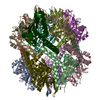
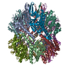
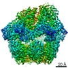
| Title | cytochrome c6 from Arthrospira maxima: an assembly of 24 subunits in the form of an oblate shell | |||||||||
 Components Components | cytochrome c6 | |||||||||
 Keywords Keywords | ELECTRON TRANSPORT / cytochrome oligomer | |||||||||
| Function / homology |  Function and homology information Function and homology informationplasma membrane-derived thylakoid lumen / photosynthesis / electron transfer activity / iron ion binding / heme binding Similarity search - Function | |||||||||
| Biological species |  Arthrospira maxima (bacteria) Arthrospira maxima (bacteria) | |||||||||
| Method |  X-RAY DIFFRACTION / SYNCHROTRON / MOLECULAR REPLACEMENT / Resolution: 3.5 Å X-RAY DIFFRACTION / SYNCHROTRON / MOLECULAR REPLACEMENT / Resolution: 3.5 Å | |||||||||
 Authors Authors | Kerfeld, C.A. / Sawaya, M.R. / Krogmann, D. / Yeates, T.O. | |||||||||
 Citation Citation | Journal: Acta Crystallogr.,Sect.D / Year: 2002 Title: Structure of cytochrome c6 from Arthrospira maxima: an assembly of 24 subunits in a nearly symmetric shell. Authors: Kerfeld, C.A. / Sawaya, M.R. / Krogmann, D.W. / Yeates, T.O. #1: Journal: Biochemistry / Year: 2001Title: Structures of Cytochrome c-549 and Cytochrome c6 from the Cyanobacterium Arthrospira maxima Authors: Sawaya, M.R. / Krogmann, D.W. / Serag, A. / Ho, K.K. / Yeates, T.O. / Kerfeld, C.A. | |||||||||
| History |
|
- Structure visualization
| Structure viewer | Molecule: MolmilJmol/JSmol |
|---|
- Downloads & links
-Download
| PDBx/mmCIF format | 1kib.cif.gz | 145.1 KB | Display | PDBx/mmCIF format |
|---|---|---|---|---|
| PDB format | pdb1kib.ent.gz | 119.2 KB | Display | PDB format |
| PDBx/mmJSON format | 1kib.json.gz | Tree view | PDBx/mmJSON format | |
| Others |  Other downloads Other downloads |
-Validation report
| Summary document | 1kib_validation.pdf.gz | 2.7 MB | Display | wwPDB validaton report |
|---|---|---|---|---|
| Full document | 1kib_full_validation.pdf.gz | 2.7 MB | Display | |
| Data in XML | 1kib_validation.xml.gz | 24.6 KB | Display | |
| Data in CIF | 1kib_validation.cif.gz | 34.9 KB | Display | |
| Arichive directory | https://data.pdbj.org/pub/pdb/validation_reports/ki/1kibftp://data.pdbj.org/pub/pdb/validation_reports/ki/1kib | HTTPS FTP |
-Related structure data
| Related structure data |  1f1fS S: Starting model for refinement |
|---|---|
| Similar structure data |


-Links
 PDBj
PDBj


- Assembly
| Deposited unit | 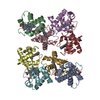
| ||||||||
|---|---|---|---|---|---|---|---|---|---|
| 1 | 
| ||||||||
| Unit cell |
|
-Components
| #1: Protein | Mass: 9246.372 Da / Num. of mol.: 8 / Source method: isolated from a natural source / Source: (natural) Arthrospira maxima (bacteria) / References: UniProt: P00118#2: Chemical | ChemComp-HEC /   Mass: 618.503 Da / Num. of mol.: 8 / Source method: obtained synthetically / Formula: C34H34FeN4O4 Mass: 618.503 Da / Num. of mol.: 8 / Source method: obtained synthetically / Formula: C34H34FeN4O4Has protein modification | Y | |
|---|
-Experimental details
-Experiment
| Experiment | Method: X-RAY DIFFRACTION / Number of used crystals: 1 |
|---|
- Sample preparation
| Crystal | Density Matthews: 3.75 Å3/Da / Density % sol: 67.19 % | |||||||||||||||||||||||||||||||||||||||||||||||||
|---|---|---|---|---|---|---|---|---|---|---|---|---|---|---|---|---|---|---|---|---|---|---|---|---|---|---|---|---|---|---|---|---|---|---|---|---|---|---|---|---|---|---|---|---|---|---|---|---|---|---|
| Crystal grow | Temperature: 298 K / Method: vapor diffusion, hanging drop / pH: 7.8 Details: 0.1M Tris pH 7.8, 2.4M ammonium sulfate, 5% methylpentanediol, VAPOR DIFFUSION, HANGING DROP, temperature 298.0K | |||||||||||||||||||||||||||||||||||||||||||||||||
| Crystal grow | *PLUS pH: 7.5 / Method: vapor diffusion | |||||||||||||||||||||||||||||||||||||||||||||||||
| Components of the solutions | *PLUS
|
-Data collection
| Diffraction | Mean temperature: 298 K |
|---|---|
| Diffraction source | Source: SYNCHROTRON / Site: NSLS  / Beamline: X8C / Wavelength: 1 Å / Beamline: X8C / Wavelength: 1 Å |
| Detector | Type: MARRESEARCH / Detector: CCD / Date: Nov 27, 1995 |
| Radiation | Monochromator: mirror / Protocol: SINGLE WAVELENGTH / Monochromatic (M) / Laue (L): M / Scattering type: x-ray |
| Radiation wavelength | Wavelength: 1 Å / Relative weight: 1 |
| Reflection | Resolution: 3.5→20 Å / Num. all: 13268 / Num. obs: 13268 / % possible obs: 91 % / Observed criterion σ(F): 0 / Observed criterion σ(I): 0 / Redundancy: 7.1 % / Rsym value: 0.15 / Net I/σ(I): 9.3 |
| Reflection shell | Resolution: 3.5→3.62 Å / Redundancy: 0.391 % / Mean I/σ(I) obs: 3.4 / Num. unique all: 1352 / % possible all: 95.5 |
| Reflection | *PLUS % possible obs: 91 % / Rmerge(I) obs: 0.15 |
| Reflection shell | *PLUS % possible obs: 95.5 % / Redundancy: 7 % / Rmerge(I) obs: 0.391 |
- Processing
| Software |
| |||||||||||||||||||||||||
|---|---|---|---|---|---|---|---|---|---|---|---|---|---|---|---|---|---|---|---|---|---|---|---|---|---|---|
| Refinement | Method to determine structure: MOLECULAR REPLACEMENT Starting model: 1f1f Resolution: 3.5→20 Å / Isotropic thermal model: isotropic / Cross valid method: THROUGHOUT / σ(F): 0 / σ(I): 0 / Stereochemistry target values: Engh & Huber
| |||||||||||||||||||||||||
| Displacement parameters | Biso mean: 41.3 Å2 | |||||||||||||||||||||||||
| Refinement step | Cycle: LAST / Resolution: 3.5→20 Å
| |||||||||||||||||||||||||
| Refine LS restraints |
| |||||||||||||||||||||||||
| Refinement | *PLUS Lowest resolution: 20 Å / % reflection Rfree: 5 % / Rfactor obs: 0.2051 / Rfactor Rfree: 0.223 / Rfactor Rwork: 0.202 | |||||||||||||||||||||||||
| Solvent computation | *PLUS | |||||||||||||||||||||||||
| Displacement parameters | *PLUS | |||||||||||||||||||||||||
| Refine LS restraints | *PLUS
|
