| 登録情報 | データベース: PDB / ID: 1ki0
|
|---|
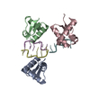
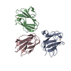
| タイトル | The X-ray Structure of Human Angiostatin |
|---|
 要素 要素 | ANGIOSTATIN |
|---|
 キーワード キーワード | HYDROLASE / kringle domains |
|---|
| 機能・相同性 |  機能・相同性情報 機能・相同性情報
plasmin / tissue remodeling / protein antigen binding / Signaling by PDGF / positive regulation of fibrinolysis / negative regulation of cell-cell adhesion mediated by cadherin / Dissolution of Fibrin Clot / biological process involved in interaction with symbiont / Activation of Matrix Metalloproteinases / apolipoprotein binding ...plasmin / tissue remodeling / protein antigen binding / Signaling by PDGF / positive regulation of fibrinolysis / negative regulation of cell-cell adhesion mediated by cadherin / Dissolution of Fibrin Clot / biological process involved in interaction with symbiont / Activation of Matrix Metalloproteinases / apolipoprotein binding / extracellular matrix disassembly / negative regulation of cell-substrate adhesion / positive regulation of blood vessel endothelial cell migration / negative regulation of fibrinolysis / fibrinolysis / Degradation of the extracellular matrix / serine-type peptidase activity / platelet alpha granule lumen / kinase binding / Regulation of Insulin-like Growth Factor (IGF) transport and uptake by Insulin-like Growth Factor Binding Proteins (IGFBPs) / blood coagulation / Platelet degranulation / protein-folding chaperone binding / protease binding / : / endopeptidase activity / blood microparticle / protein domain specific binding / external side of plasma membrane / signaling receptor binding / negative regulation of cell population proliferation / serine-type endopeptidase activity / enzyme binding / cell surface / proteolysis / extracellular space / extracellular exosome / extracellular region / plasma membrane類似検索 - 分子機能 Peptidase S1A, plasmin / Plasminogen Kringle 4 / Plasminogen Kringle 4 / divergent subfamily of APPLE domains / : / PAN/Apple domain profile. / PAN domain / PAN/Apple domain / Kringle domain / Kringle ...Peptidase S1A, plasmin / Plasminogen Kringle 4 / Plasminogen Kringle 4 / divergent subfamily of APPLE domains / : / PAN/Apple domain profile. / PAN domain / PAN/Apple domain / Kringle domain / Kringle / Kringle, conserved site / Kringle superfamily / Kringle domain signature. / Kringle domain profile. / Kringle domain / Kringle-like fold / Serine proteases, trypsin family, histidine active site / Serine proteases, trypsin family, serine active site / Serine proteases, trypsin family, histidine active site. / Peptidase S1A, chymotrypsin family / Serine proteases, trypsin family, serine active site. / Serine proteases, trypsin domain profile. / Trypsin-like serine protease / Serine proteases, trypsin domain / Trypsin / Peptidase S1, PA clan, chymotrypsin-like fold / Peptidase S1, PA clan / Beta Barrel / Mainly Beta類似検索 - ドメイン・相同性 |
|---|
| 生物種 |  Homo sapiens (ヒト) Homo sapiens (ヒト) |
|---|
| 手法 |  X線回折 / シンクロトロン / 分子置換 / 解像度: 1.75 Å X線回折 / シンクロトロン / 分子置換 / 解像度: 1.75 Å |
|---|
 データ登録者 データ登録者 | Abad, M.C. / Arni, R.K. / Grella, D.K. / Castellino, F.J. / Tulinsky, A. / Geiger, J.H. |
|---|
 引用 引用 | ジャーナル: J.Mol.Biol. / 年: 2002
タイトル: The X-ray crystallographic structure of the angiogenesis inhibitor angiostatin.
著者: Abad, M.C. / Arni, R.K. / Grella, D.K. / Castellino, F.J. / Tulinsky, A. / Geiger, J.H. |
|---|
| 履歴 | | 登録 | 2001年12月2日 | 登録サイト: RCSB / 処理サイト: RCSB |
|---|
| 改定 1.0 | 2002年5月29日 | Provider: repository / タイプ: Initial release |
|---|
| 改定 1.1 | 2008年4月27日 | Group: Version format compliance |
|---|
| 改定 1.2 | 2011年7月13日 | Group: Version format compliance |
|---|
| 改定 1.3 | 2021年10月27日 | Group: Database references / Derived calculations / カテゴリ: database_2 / struct_ref_seq_dif / struct_site
Item: _database_2.pdbx_DOI / _database_2.pdbx_database_accession ..._database_2.pdbx_DOI / _database_2.pdbx_database_accession / _struct_ref_seq_dif.details / _struct_site.pdbx_auth_asym_id / _struct_site.pdbx_auth_comp_id / _struct_site.pdbx_auth_seq_id |
|---|
| 改定 1.4 | 2024年11月6日 | Group: Data collection / Structure summary
カテゴリ: chem_comp_atom / chem_comp_bond ...chem_comp_atom / chem_comp_bond / pdbx_entry_details / pdbx_modification_feature |
|---|
|
|---|
 ムービー
ムービー コントローラー


 データを開く
 基本情報
基本情報 構造の表示
構造の表示 ダウンロードとリンク
 その他のダウンロード


 PDBj
PDBj


 集合体
集合体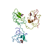
 Pichia pastoris (菌類) / 参照: UniProt: P00747, plasmin
 分子量: 163.172 Da / 分子数: 3 / 由来タイプ: 合成 / 式: C6H13NO4 / コメント: pH緩衝剤*YM
 分子量: 18.015 Da / 分子数: 393 / 由来タイプ: 天然 / 式: H2O
 試料調製
試料調製 / ビームライン: 19-ID / 波長: 1.0332 Å
 解析
解析