 Movie
Movie Controller

[English] 日本語
 Yorodumi
- PDB-1kh0: Accurate Computer Base Design of a New Backbone Conformation in t... -
+ Open data

- Basic information
| Entry | Database: PDB / ID: 1kh0 | ||||||
|---|---|---|---|---|---|---|---|
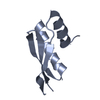
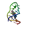
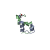
| Title | Accurate Computer Base Design of a New Backbone Conformation in the Second Turn of Protein L | ||||||
 Components Components | protein L | ||||||
 Keywords Keywords | PROTEIN BINDING / Protein L B1 domain / computational based protein design / Type 1' beta turn / extensive amino acid mutations. | ||||||
| Function / homology |  Function and homology information Function and homology information | ||||||
| Biological species |  Finegoldia magna (bacteria) Finegoldia magna (bacteria) | ||||||
| Method |  X-RAY DIFFRACTION / MOLECULAR REPLACEMENT / Resolution: 1.9 Å X-RAY DIFFRACTION / MOLECULAR REPLACEMENT / Resolution: 1.9 Å | ||||||
 Authors Authors | O'Neill, J.W. / Kuhlman, B. / Kim, D.E. / Zhang, K.Y. / Baker, D. | ||||||
 Citation Citation | Journal: J.Mol.Biol. / Year: 2002 Title: Accurate computer-based design of a new backbone conformation in the second turn of protein L. Authors: Kuhlman, B. / O'Neill, J.W. / Kim, D.E. / Zhang, K.Y. / Baker, D. | ||||||
| History |
|
- Structure visualization
| Structure viewer | Molecule: MolmilJmol/JSmol |
|---|
- Downloads & links
-Download
| PDBx/mmCIF format | 1kh0.cif.gz | 37.7 KB | Display | PDBx/mmCIF format |
|---|---|---|---|---|
| PDB format | pdb1kh0.ent.gz | 26 KB | Display | PDB format |
| PDBx/mmJSON format | 1kh0.json.gz | Tree view | PDBx/mmJSON format | |
| Others |  Other downloads Other downloads |
-Validation report
| Arichive directory | https://data.pdbj.org/pub/pdb/validation_reports/kh/1kh0ftp://data.pdbj.org/pub/pdb/validation_reports/kh/1kh0 | HTTPS FTP |
|---|
-Related structure data
| Related structure data |  1hz6S S: Starting model for refinement |
|---|---|
| Similar structure data |


-Links
 PDBj
PDBj- Assembly
| Deposited unit | 
| ||||||||
|---|---|---|---|---|---|---|---|---|---|
| 1 | 
| ||||||||
| 2 | 
| ||||||||
| Unit cell |
|
-Components
| #1: Protein | Mass: 7124.085 Da / Num. of mol.: 2 / Fragment: B1 DOMAIN (RESIDUES 111-173) Mutation: F26K, T30L, A33V, Y34L, Y47W, V49I, A54T, D55N, K56G, G57B, Y58I, T59I Source method: isolated from a genetically manipulated source Source: (gene. exp.) Finegoldia magna (bacteria) / Strain: ATCC 29328 / Plasmid: pET3A / Species (production host): Escherichia coli / Production host: #2: Water | ChemComp-HOH / |  Mass: 18.015 Da / Num. of mol.: 80 / Source method: isolated from a natural source / Formula: H2O Mass: 18.015 Da / Num. of mol.: 80 / Source method: isolated from a natural source / Formula: H2O |
|---|
-Experimental details
-Experiment
| Experiment | Method: X-RAY DIFFRACTION / Number of used crystals: 1 |
|---|
- Sample preparation
| Crystal | Density Matthews: 2.2 Å3/Da / Density % sol: 43.74 % | ||||||||||||||||||||||||||||||||||||||||||
|---|---|---|---|---|---|---|---|---|---|---|---|---|---|---|---|---|---|---|---|---|---|---|---|---|---|---|---|---|---|---|---|---|---|---|---|---|---|---|---|---|---|---|---|
| Crystal grow | Temperature: 277 K / Method: vapor diffusion, hanging drop / pH: 6.5 Details: 1M Sodium Citrate, 100mM Cacodylate, pH 6.5, VAPOR DIFFUSION, HANGING DROP, temperature 277K | ||||||||||||||||||||||||||||||||||||||||||
| Crystal grow | *PLUS | ||||||||||||||||||||||||||||||||||||||||||
| Components of the solutions | *PLUS
|
-Data collection
| Diffraction | Mean temperature: 100 K |
|---|---|
| Diffraction source | Source: ROTATING ANODE / Type: RIGAKU / Wavelength: 1.5418 Å |
| Detector | Type: RIGAKU RAXIS IV / Detector: IMAGE PLATE / Date: Oct 22, 2000 / Details: mirrors |
| Radiation | Monochromator: Yale Mirrors / Protocol: SINGLE WAVELENGTH / Monochromatic (M) / Laue (L): M / Scattering type: x-ray |
| Radiation wavelength | Wavelength: 1.5418 Å / Relative weight: 1 |
| Reflection | Resolution: 1.9→25 Å / Num. all: 11610 / Num. obs: 11610 / % possible obs: 96.9 % / Observed criterion σ(F): -3 / Observed criterion σ(I): -3 / Redundancy: 6.9 % / Biso Wilson estimate: 23.4 Å2 / Rmerge(I) obs: 0.049 / Rsym value: 0.043 / Net I/σ(I): 36.4 |
| Reflection shell | Resolution: 1.9→1.96 Å / Redundancy: 6.8 % / Rmerge(I) obs: 0.27 / Mean I/σ(I) obs: 6.9 / Num. unique all: 923 / Rsym value: 0.252 / % possible all: 95.1 |
| Reflection | *PLUS Lowest resolution: 25 Å |
- Processing
| Software |
| ||||||||||||||||||||||||||||||||||||||||||||||||||||||||||||||||||||||||||||||||
|---|---|---|---|---|---|---|---|---|---|---|---|---|---|---|---|---|---|---|---|---|---|---|---|---|---|---|---|---|---|---|---|---|---|---|---|---|---|---|---|---|---|---|---|---|---|---|---|---|---|---|---|---|---|---|---|---|---|---|---|---|---|---|---|---|---|---|---|---|---|---|---|---|---|---|---|---|---|---|---|---|---|
| Refinement | Method to determine structure: MOLECULAR REPLACEMENT Starting model: PDB ENTRY 1HZ6 Resolution: 1.9→23.09 Å / Rfactor Rfree error: 0.007 / Data cutoff high absF: 1027037.39 / Data cutoff low absF: 0 / Isotropic thermal model: RESTRAINED / Cross valid method: THROUGHOUT / σ(F): 0 / σ(I): 0 / Stereochemistry target values: Maximum likelihood
| ||||||||||||||||||||||||||||||||||||||||||||||||||||||||||||||||||||||||||||||||
| Solvent computation | Solvent model: FLAT MODEL / Bsol: 54.2031 Å2 / ksol: 0.341182 e/Å3 | ||||||||||||||||||||||||||||||||||||||||||||||||||||||||||||||||||||||||||||||||
| Displacement parameters | Biso mean: 34.9 Å2
| ||||||||||||||||||||||||||||||||||||||||||||||||||||||||||||||||||||||||||||||||
| Refine analyze |
| ||||||||||||||||||||||||||||||||||||||||||||||||||||||||||||||||||||||||||||||||
| Refinement step | Cycle: LAST / Resolution: 1.9→23.09 Å
| ||||||||||||||||||||||||||||||||||||||||||||||||||||||||||||||||||||||||||||||||
| Refine LS restraints |
| ||||||||||||||||||||||||||||||||||||||||||||||||||||||||||||||||||||||||||||||||
| LS refinement shell | Resolution: 1.9→2.02 Å / Rfactor Rfree error: 0.02 / Total num. of bins used: 6
| ||||||||||||||||||||||||||||||||||||||||||||||||||||||||||||||||||||||||||||||||
| Xplor file |
| ||||||||||||||||||||||||||||||||||||||||||||||||||||||||||||||||||||||||||||||||
| Software | *PLUS Name: CNS / Version: 1 / Classification: refinement | ||||||||||||||||||||||||||||||||||||||||||||||||||||||||||||||||||||||||||||||||
| Refinement | *PLUS σ(F): 0 / % reflection Rfree: 9.8 % | ||||||||||||||||||||||||||||||||||||||||||||||||||||||||||||||||||||||||||||||||
| Solvent computation | *PLUS | ||||||||||||||||||||||||||||||||||||||||||||||||||||||||||||||||||||||||||||||||
| Displacement parameters | *PLUS Biso mean: 34.9 Å2 | ||||||||||||||||||||||||||||||||||||||||||||||||||||||||||||||||||||||||||||||||
| Refine LS restraints | *PLUS
| ||||||||||||||||||||||||||||||||||||||||||||||||||||||||||||||||||||||||||||||||
| LS refinement shell | *PLUS Rfactor Rfree: 0.265 / % reflection Rfree: 9.2 % / Rfactor Rwork: 0.208 |
