 Movie
Movie Controller

[English] 日本語
 Yorodumi
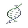
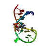
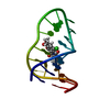
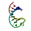
- PDB-1ecu: SOLUTION STRUCTURE OF E2F BINDING DNA FRAGMENT GCGCGAAAC-T-GTTTCGCGC -
+ Open data

- Basic information
| Entry | Database: PDB / ID: 1ecu | ||||||||||||||||||
|---|---|---|---|---|---|---|---|---|---|---|---|---|---|---|---|---|---|---|---|
| Title | SOLUTION STRUCTURE OF E2F BINDING DNA FRAGMENT GCGCGAAAC-T-GTTTCGCGC | ||||||||||||||||||
 Components Components | DNA (5'-D(* Keywords KeywordsDNA / double strands / loop | Function / homology | DNA / DNA (> 10) |  Function and homology information Function and homology informationMethod | SOLUTION NMR / Molecular Dynamics with Particle-Particle Particle-Mesh method; Iterative Relaxation Matrix Approach with generalized order parameters |  Authors AuthorsWu, J.H. / Chang, C. / Pei, J.M. / Xiao, Q. / Shi, Y.Y. |  CitationJournal: to be published / Year: 2000 CitationJournal: to be published / Year: 2000Title: Solution structure of E2F binding DNA fragment GCGCGAAAC-T-GTTTCGCGC studied by Molecular Dynamics Simulation and Two Dimensional NMR experiment Authors: Wu, J.H. / Chang, C. / Pei, J.M. / Xiao, Q. / Shi, Y.Y. History |
|
- Structure visualization
| Structure viewer | Molecule: MolmilJmol/JSmol |
|---|
- Downloads & links
-Download
| PDBx/mmCIF format | 1ecu.cif.gz | 29.5 KB | Display | PDBx/mmCIF format |
|---|---|---|---|---|
| PDB format | pdb1ecu.ent.gz | 17.8 KB | Display | PDB format |
| PDBx/mmJSON format | 1ecu.json.gz | Tree view | PDBx/mmJSON format | |
| Others |  Other downloads Other downloads |
-Validation report
| Arichive directory | https://data.pdbj.org/pub/pdb/validation_reports/ec/1ecuftp://data.pdbj.org/pub/pdb/validation_reports/ec/1ecu | HTTPS FTP |
|---|
-Related structure data
| Similar structure data |
|---|


-Links
 PDBj
PDBj


- Assembly
| Deposited unit | 
| |||||||||
|---|---|---|---|---|---|---|---|---|---|---|
| 1 |
| |||||||||
| NMR ensembles |
|
-Components
| #1: DNA chain | Mass: 5821.759 Da / Num. of mol.: 1 / Fragment: FRAGMENT OF E2F BINDING DNA / Source method: obtained synthetically Details: The sequence is formed by adding CTG loop to ends of ds(GCGCGAAA:TTTCGCGC), which occurs naturally in humans. |
|---|
-Experimental details
-Experiment
| Experiment | Method: SOLUTION NMR | ||||||||||||
|---|---|---|---|---|---|---|---|---|---|---|---|---|---|
| NMR experiment |
| ||||||||||||
| NMR details | Text: Solvent suppression was realized by WATERGATE method. |
- Sample preparation
| Details |
| |||||||||
|---|---|---|---|---|---|---|---|---|---|---|
| Sample conditions | Ionic strength: 150mM NaCl / pH: 7 / Pressure: ambient / Temperature: 300 K |
-NMR measurement
| NMR spectrometer | Type: Bruker DMX / Manufacturer: Bruker / Model: DMX / Field strength: 500 MHz |
|---|
- Processing
| NMR software |
| ||||||||||||||||
|---|---|---|---|---|---|---|---|---|---|---|---|---|---|---|---|---|---|
| Refinement | Method: Molecular Dynamics with Particle-Particle Particle-Mesh method; Iterative Relaxation Matrix Approach with generalized order parameters Software ordinal: 1 Details: Initial structure for model 1 is A-DNA, while that for model 2 is B-DNA. First we applied 640ps free MD with 18 Na+ counterions and 2789 waters for A-DNA and 2303 waters for B-DNA. Then we ...Details: Initial structure for model 1 is A-DNA, while that for model 2 is B-DNA. First we applied 640ps free MD with 18 Na+ counterions and 2789 waters for A-DNA and 2303 waters for B-DNA. Then we applied 4 cycles of IRMA with 174 NOE restraints and 19 hydrogen bond restraints | ||||||||||||||||
| NMR ensemble | Conformer selection criteria: all calculated structures submitted Conformers calculated total number: 2 / Conformers submitted total number: 2 |
