 Movie
Movie Controller


+ Open data

- Basic information
| Entry | Database: PDB / ID: 1keh | ||||||
|---|---|---|---|---|---|---|---|
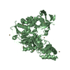
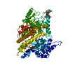
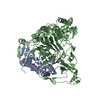
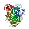
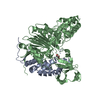
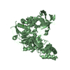
| Title | Precursor structure of cephalosporin acylase | ||||||
 Components Components | precursor of cephalosporin acylase | ||||||
 Keywords Keywords | HYDROLASE / cephalosporin acylase / precursor / glutaryl-7-ACA | ||||||
| Function / homology |  Function and homology information Function and homology informationglutaryl-7-aminocephalosporanic-acid acylase / glutaryl-7-aminocephalosporanic-acid acylase activity / antibiotic biosynthetic process / periplasmic space / response to antibiotic Similarity search - Function | ||||||
| Biological species |  Brevundimonas diminuta (bacteria) Brevundimonas diminuta (bacteria) | ||||||
| Method |  X-RAY DIFFRACTION / SYNCHROTRON / FOURIER SYNTHESIS / Resolution: 2.5 Å X-RAY DIFFRACTION / SYNCHROTRON / FOURIER SYNTHESIS / Resolution: 2.5 Å | ||||||
 Authors Authors | Kim, Y. / Kim, S. | ||||||
 Citation Citation | Journal: J.Biol.Chem. / Year: 2002 Title: Precursor structure of cephalosporin acylase. Insights into autoproteolytic activation in a new N-terminal hydrolase family Authors: Kim, Y. / Kim, S. / Earnest, T.N. / Hol, W.G. | ||||||
| History |
|
- Structure visualization
| Structure viewer | Molecule: MolmilJmol/JSmol |
|---|
- Downloads & links
-Download
| PDBx/mmCIF format | 1keh.cif.gz | 151.3 KB | Display | PDBx/mmCIF format |
|---|---|---|---|---|
| PDB format | pdb1keh.ent.gz | 118.4 KB | Display | PDB format |
| PDBx/mmJSON format | 1keh.json.gz | Tree view | PDBx/mmJSON format | |
| Others |  Other downloads Other downloads |
-Validation report
| Arichive directory | https://data.pdbj.org/pub/pdb/validation_reports/ke/1kehftp://data.pdbj.org/pub/pdb/validation_reports/ke/1keh | HTTPS FTP |
|---|
-Related structure data
| Similar structure data |
|---|
-Links
 PDBj
PDBj


- Assembly
| Deposited unit | 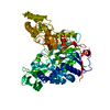
| ||||||||
|---|---|---|---|---|---|---|---|---|---|
| 1 |
| ||||||||
| Unit cell |
|
-Components
| #1: Protein | Mass: 76609.883 Da / Num. of mol.: 1 / Fragment: residues 1-689 / Mutation: S170A,R428A Source method: isolated from a genetically manipulated source Source: (gene. exp.) Brevundimonas diminuta (bacteria) / Plasmid: pET24d(+) / Production host: References: UniProt: Q9L5D6, Hydrolases; Acting on carbon-nitrogen bonds, other than peptide bonds; In linear amides |
|---|---|
| #2: Water | ChemComp-HOH /  Mass: 18.015 Da / Num. of mol.: 399 / Source method: isolated from a natural source / Formula: H2O Mass: 18.015 Da / Num. of mol.: 399 / Source method: isolated from a natural source / Formula: H2O |
-Experimental details
-Experiment
| Experiment | Method: X-RAY DIFFRACTION / Number of used crystals: 1 |
|---|
- Sample preparation
| Crystal | Density Matthews: 3.37 Å3/Da / Density % sol: 63.55 % | ||||||||||||||||||||||||||||||||||||||||||||||||||||||||
|---|---|---|---|---|---|---|---|---|---|---|---|---|---|---|---|---|---|---|---|---|---|---|---|---|---|---|---|---|---|---|---|---|---|---|---|---|---|---|---|---|---|---|---|---|---|---|---|---|---|---|---|---|---|---|---|---|---|
| Crystal grow | Temperature: 290 K / Method: vapor diffusion, hanging drop / pH: 6.5 Details: PEG8000, MgAcetate, Sodium Cacodylate, DTT, pH 6.5, VAPOR DIFFUSION, HANGING DROP, temperature 290K | ||||||||||||||||||||||||||||||||||||||||||||||||||||||||
| Crystal grow | *PLUS Temperature: 21 ℃ / pH: 7 | ||||||||||||||||||||||||||||||||||||||||||||||||||||||||
| Components of the solutions | *PLUS
|
-Data collection
| Diffraction | Mean temperature: 125 K |
|---|---|
| Diffraction source | Source: SYNCHROTRON / Site: ALS  / Beamline: 5.0.2 / Wavelength: 0.91 Å / Beamline: 5.0.2 / Wavelength: 0.91 Å |
| Detector | Type: CUSTOM-MADE / Detector: CCD / Date: Jan 7, 2001 |
| Radiation | Monochromator: graphite / Protocol: SINGLE WAVELENGTH / Monochromatic (M) / Laue (L): M / Scattering type: x-ray |
| Radiation wavelength | Wavelength: 0.91 Å / Relative weight: 1 |
| Reflection | Resolution: 2.5→20 Å / Num. all: 39792 / Num. obs: 38001 / % possible obs: 95.5 % / Observed criterion σ(F): 0 / Observed criterion σ(I): 3.2 |
| Reflection shell | Resolution: 2.5→2.59 Å / % possible all: 70.4 |
| Reflection | *PLUS Lowest resolution: 20 Å / Num. measured all: 838095 / Rmerge(I) obs: 0.108 |
| Reflection shell | *PLUS Highest resolution: 2.5 Å / % possible obs: 70.4 % / Rmerge(I) obs: 0.548 / Mean I/σ(I) obs: 3.2 |
- Processing
| Software |
| |||||||||||||||||||||||||
|---|---|---|---|---|---|---|---|---|---|---|---|---|---|---|---|---|---|---|---|---|---|---|---|---|---|---|
| Refinement | Method to determine structure: FOURIER SYNTHESIS / Resolution: 2.5→20 Å / σ(F): 0 / Stereochemistry target values: Engh & Huber
| |||||||||||||||||||||||||
| Refinement step | Cycle: LAST / Resolution: 2.5→20 Å
| |||||||||||||||||||||||||
| Refinement | *PLUS Lowest resolution: 20 Å / % reflection Rfree: 3.5 % / Rfactor obs: 0.203 | |||||||||||||||||||||||||
| Solvent computation | *PLUS | |||||||||||||||||||||||||
| Displacement parameters | *PLUS | |||||||||||||||||||||||||
| Refine LS restraints | *PLUS
|
