 Movie
Movie Controller


+ Open data

- Basic information
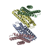
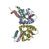
| Entry | Database: PDB / ID: 1kaw | ||||||
|---|---|---|---|---|---|---|---|
| Title | STRUCTURE OF SINGLE STRANDED DNA BINDING PROTEIN (SSB) | ||||||
 Components Components | SINGLE-STRANDED DNA BINDING PROTEIN | ||||||
 Keywords Keywords | DNA BINDING PROTEIN / DNA-BINDING PROTEIN / SINGLE STRANDED DNA BINDING PROTEIN / SSB | ||||||
| Function / homology |  Function and homology information Function and homology informationsingle-stranded DNA-binding protein complex / nucleoid / SOS response / replisome / recombinational repair / mismatch repair / DNA-templated DNA replication / enzyme activator activity / single-stranded DNA binding / DNA recombination ...single-stranded DNA-binding protein complex / nucleoid / SOS response / replisome / recombinational repair / mismatch repair / DNA-templated DNA replication / enzyme activator activity / single-stranded DNA binding / DNA recombination / DNA replication / DNA repair / identical protein binding / cytosol Similarity search - Function | ||||||
| Biological species |  | ||||||
| Method |  X-RAY DIFFRACTION / SYNCHROTRON / Resolution: 2.9 Å X-RAY DIFFRACTION / SYNCHROTRON / Resolution: 2.9 Å | ||||||
 Authors Authors | Raghunathan, S. / Waksman, G. | ||||||
 Citation Citation | Journal: Proc.Natl.Acad.Sci.USA / Year: 1997 Title: Crystal structure of the homo-tetrameric DNA binding domain of Escherichia coli single-stranded DNA-binding protein determined by multiwavelength x-ray diffraction on the selenomethionyl ...Title: Crystal structure of the homo-tetrameric DNA binding domain of Escherichia coli single-stranded DNA-binding protein determined by multiwavelength x-ray diffraction on the selenomethionyl protein at 2.9-A resolution. Authors: Raghunathan, S. / Ricard, C.S. / Lohman, T.M. / Waksman, G. | ||||||
| History |
|
- Structure visualization
| Structure viewer | Molecule: MolmilJmol/JSmol |
|---|
- Downloads & links
-Download
| PDBx/mmCIF format | 1kaw.cif.gz | 86.2 KB | Display | PDBx/mmCIF format |
|---|---|---|---|---|
| PDB format | pdb1kaw.ent.gz | 66.7 KB | Display | PDB format |
| PDBx/mmJSON format | 1kaw.json.gz | Tree view | PDBx/mmJSON format | |
| Others |  Other downloads Other downloads |
-Validation report
| Arichive directory | https://data.pdbj.org/pub/pdb/validation_reports/ka/1kawftp://data.pdbj.org/pub/pdb/validation_reports/ka/1kaw | HTTPS FTP |
|---|
-Related structure data
| Similar structure data |
|---|


-Links
 PDBj
PDBj- Assembly
| Deposited unit | 
| ||||||||||||
|---|---|---|---|---|---|---|---|---|---|---|---|---|---|
| 1 |
| ||||||||||||
| Unit cell |
| ||||||||||||
| Noncrystallographic symmetry (NCS) | NCS oper:
|
-Components
| #1: Protein | Mass: 14480.156 Da / Num. of mol.: 4 / Fragment: CHYMOTRYPTIC FRAGMENT, RESIDUES 1 - 135 / Source method: isolated from a natural source / Source: (natural) #2: Water | ChemComp-HOH / |  Mass: 18.015 Da / Num. of mol.: 35 / Source method: isolated from a natural source / Formula: H2O Mass: 18.015 Da / Num. of mol.: 35 / Source method: isolated from a natural source / Formula: H2O |
|---|
-Experimental details
-Experiment
| Experiment | Method: X-RAY DIFFRACTION / Number of used crystals: 1 |
|---|
- Sample preparation
| Crystal | Density Matthews: 2.36 Å3/Da / Density % sol: 52 % | |||||||||||||||
|---|---|---|---|---|---|---|---|---|---|---|---|---|---|---|---|---|
| Crystal grow | *PLUS pH: 8.5 / Method: vapor diffusion, hanging drop | |||||||||||||||
| Components of the solutions | *PLUS
|
-Data collection
| Diffraction | Mean temperature: 100 K |
|---|---|
| Diffraction source | Source: SYNCHROTRON / Site: SSRL  / Beamline: BL7-1 / Wavelength: 1.08 / Beamline: BL7-1 / Wavelength: 1.08 |
| Detector | Type: MARRESEARCH / Detector: IMAGE PLATE / Date: Nov 11, 1995 |
| Radiation | Monochromatic (M) / Laue (L): M / Scattering type: x-ray |
| Radiation wavelength | Wavelength: 1.08 Å / Relative weight: 1 |
| Reflection | Num. obs: 14346 / % possible obs: 89.7 % / Observed criterion σ(I): 1 / Redundancy: 2.6 % / Rmerge(I) obs: 0.053 |
| Reflection | *PLUS Highest resolution: 2.7 Å / Lowest resolution: 30 Å / Num. obs: 12833 / % possible obs: 83.6 % |
- Processing
| Software |
| ||||||||||||||||||||||||||||||||||||||||||||||||||||||||||||
|---|---|---|---|---|---|---|---|---|---|---|---|---|---|---|---|---|---|---|---|---|---|---|---|---|---|---|---|---|---|---|---|---|---|---|---|---|---|---|---|---|---|---|---|---|---|---|---|---|---|---|---|---|---|---|---|---|---|---|---|---|---|
| Refinement | Resolution: 2.9→8 Å / σ(F): 1 Details: MOLECULAR DYNAMICS RESTRAINED REFINEMENT FOLLOWED BY LEAST-SQUARES OPTIMIZATION
| ||||||||||||||||||||||||||||||||||||||||||||||||||||||||||||
| Displacement parameters | Biso mean: 21.9 Å2 | ||||||||||||||||||||||||||||||||||||||||||||||||||||||||||||
| Refinement step | Cycle: LAST / Resolution: 2.9→8 Å
| ||||||||||||||||||||||||||||||||||||||||||||||||||||||||||||
| Refine LS restraints |
| ||||||||||||||||||||||||||||||||||||||||||||||||||||||||||||
| Software | *PLUS Name: X-PLOR / Version: 3.85 / Classification: refinement | ||||||||||||||||||||||||||||||||||||||||||||||||||||||||||||
| Refinement | *PLUS % reflection Rfree: 10 % / Rfactor obs: 0.23 / Rfactor Rfree: 0.295 | ||||||||||||||||||||||||||||||||||||||||||||||||||||||||||||
| Solvent computation | *PLUS | ||||||||||||||||||||||||||||||||||||||||||||||||||||||||||||
| Displacement parameters | *PLUS | ||||||||||||||||||||||||||||||||||||||||||||||||||||||||||||
| Refine LS restraints | *PLUS
| ||||||||||||||||||||||||||||||||||||||||||||||||||||||||||||
| LS refinement shell | *PLUS Highest resolution: 2.9 Å / Lowest resolution: 3 Å |
