 Movie
Movie Controller

[English] 日本語
 Yorodumi
- PDB-1kam: Structure of Bacillus subtilis Nicotinic Acid Mononucleotide Aden... -
+ Open data

- Basic information
| Entry | Database: PDB / ID: 1kam | ||||||
|---|---|---|---|---|---|---|---|
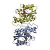
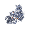
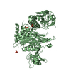
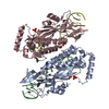
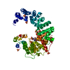
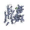
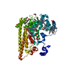
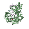
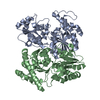
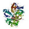
| Title | Structure of Bacillus subtilis Nicotinic Acid Mononucleotide Adenylyl Transferase | ||||||
 Components Components | NICOTINATE-NUCLEOTIDE ADENYLYLTRANSFERASE | ||||||
 Keywords Keywords | TRANSFERASE / Rossmann fold | ||||||
| Function / homology |  Function and homology information Function and homology informationnicotinate-nucleotide adenylyltransferase / nicotinate-nucleotide adenylyltransferase activity / NAD+ biosynthetic process / ATP binding Similarity search - Function | ||||||
| Biological species |  | ||||||
| Method |  X-RAY DIFFRACTION / SYNCHROTRON / MAD / Resolution: 2.1 Å X-RAY DIFFRACTION / SYNCHROTRON / MAD / Resolution: 2.1 Å | ||||||
 Authors Authors | Olland, A.M. / Underwood, K.W. / Czerwinski, R.M. / Lo, M.C. / Aulabaugh, A. / Bard, J. / Stahl, M.L. / Somers, W.S. / Sullivan, F.X. / Chopra, R. | ||||||
 Citation Citation | Journal: J.Biol.Chem. / Year: 2002 Title: Identification, characterization, and crystal structure of Bacillus subtilis nicotinic acid mononucleotide adenylyltransferase. Authors: Olland, A.M. / Underwood, K.W. / Czerwinski, R.M. / Lo, M.C. / Aulabaugh, A. / Bard, J. / Stahl, M.L. / Somers, W.S. / Sullivan, F.X. / Chopra, R. | ||||||
| History |
|
- Structure visualization
| Structure viewer | Molecule: MolmilJmol/JSmol |
|---|
- Downloads & links
-Download
| PDBx/mmCIF format | 1kam.cif.gz | 152.3 KB | Display | PDBx/mmCIF format |
|---|---|---|---|---|
| PDB format | pdb1kam.ent.gz | 122.1 KB | Display | PDB format |
| PDBx/mmJSON format | 1kam.json.gz | Tree view | PDBx/mmJSON format | |
| Others |  Other downloads Other downloads |
-Validation report
| Summary document | 1kam_validation.pdf.gz | 392.6 KB | Display | wwPDB validaton report |
|---|---|---|---|---|
| Full document | 1kam_full_validation.pdf.gz | 409.5 KB | Display | |
| Data in XML | 1kam_validation.xml.gz | 16.7 KB | Display | |
| Data in CIF | 1kam_validation.cif.gz | 25.5 KB | Display | |
| Arichive directory | https://data.pdbj.org/pub/pdb/validation_reports/ka/1kamftp://data.pdbj.org/pub/pdb/validation_reports/ka/1kam | HTTPS FTP |
-Related structure data

-Links
 PDBj
PDBj- Assembly
| Deposited unit | 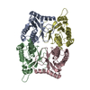
| ||||||||||
|---|---|---|---|---|---|---|---|---|---|---|---|
| 1 |
| ||||||||||
| Unit cell |
|
-Components
| #1: Protein | Mass: 22618.902 Da / Num. of mol.: 4 Source method: isolated from a genetically manipulated source Source: (gene. exp.) References: UniProt: P54455, nicotinate-nucleotide adenylyltransferase #2: Water | ChemComp-HOH / |  Mass: 18.015 Da / Num. of mol.: 139 / Source method: isolated from a natural source / Formula: H2O Mass: 18.015 Da / Num. of mol.: 139 / Source method: isolated from a natural source / Formula: H2O |
|---|
-Experimental details
-Experiment
| Experiment | Method: X-RAY DIFFRACTION / Number of used crystals: 1 |
|---|
- Sample preparation
| Crystal | Density Matthews: 2.16 Å3/Da / Density % sol: 43.03 % | ||||||||||||||||||||||||||||||||||||||||||
|---|---|---|---|---|---|---|---|---|---|---|---|---|---|---|---|---|---|---|---|---|---|---|---|---|---|---|---|---|---|---|---|---|---|---|---|---|---|---|---|---|---|---|---|
| Crystal grow | Temperature: 291 K / Method: vapor diffusion, hanging drop / pH: 7.5 Details: PEG3350, Magnesium chloride, pH 7.5, VAPOR DIFFUSION, HANGING DROP at 291K | ||||||||||||||||||||||||||||||||||||||||||
| Crystal grow | *PLUS Temperature: 18 ℃ | ||||||||||||||||||||||||||||||||||||||||||
| Components of the solutions | *PLUS
|
-Data collection
| Diffraction | Mean temperature: 93 K | |||||||||
|---|---|---|---|---|---|---|---|---|---|---|
| Diffraction source | Source: SYNCHROTRON / Site: ALS  / Beamline: 5.0.2 / Wavelength: 0.9792, 0.9567 / Beamline: 5.0.2 / Wavelength: 0.9792, 0.9567 | |||||||||
| Detector | Type: ADSC QUANTUM 4 / Detector: CCD / Date: Jul 1, 2001 | |||||||||
| Radiation | Monochromator: double crystal / Protocol: MAD / Monochromatic (M) / Laue (L): M / Scattering type: x-ray | |||||||||
| Radiation wavelength |
| |||||||||
| Reflection | Resolution: 2→20 Å / Num. all: 51680 / Num. obs: 51628 / % possible obs: 99.9 % / Observed criterion σ(I): 2 | |||||||||
| Reflection shell | Resolution: 2→2.07 Å / Rmerge(I) obs: 0.47 / Mean I/σ(I) obs: 2.4 / Num. unique all: 5108 / % possible all: 99.8 | |||||||||
| Reflection | *PLUS Highest resolution: 2 Å / Lowest resolution: 20 Å / Num. obs: 51680 / Num. measured all: 196259 / Rmerge(I) obs: 0.079 | |||||||||
| Reflection shell | *PLUS % possible obs: 99.8 % / Rmerge(I) obs: 0.47 |
- Processing
| Software |
| |||||||||||||||||||||||||
|---|---|---|---|---|---|---|---|---|---|---|---|---|---|---|---|---|---|---|---|---|---|---|---|---|---|---|
| Refinement | Method to determine structure: MAD / Resolution: 2.1→20 Å / Isotropic thermal model: isotropic / Cross valid method: THROUGHOUT / σ(F): 4 / σ(I): 2 / Stereochemistry target values: Engh & Huber
| |||||||||||||||||||||||||
| Refinement step | Cycle: LAST / Resolution: 2.1→20 Å
| |||||||||||||||||||||||||
| Refine LS restraints |
| |||||||||||||||||||||||||
| LS refinement shell | Resolution: 2.1→2.12 Å
| |||||||||||||||||||||||||
| Refinement | *PLUS Highest resolution: 2.1 Å / % reflection Rfree: 5 % / Rfactor Rfree: 0.2544 / Rfactor Rwork: 0.2221 | |||||||||||||||||||||||||
| Solvent computation | *PLUS | |||||||||||||||||||||||||
| Displacement parameters | *PLUS | |||||||||||||||||||||||||
| LS refinement shell | *PLUS Rfactor Rfree: 0.4063 / Rfactor Rwork: 0.2764 |
