+ Open data
- Basic information
| Entry | Database: PDB / ID: 1k88 | ||||||
|---|---|---|---|---|---|---|---|
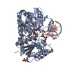
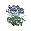
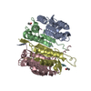
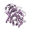
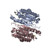
| Title | Crystal structure of procaspase-7 | ||||||
 Components Components | procaspase-7 | ||||||
 Keywords Keywords | APOPTOSIS / procaspase activation / protease / substrate binding | ||||||
| Function / homology |  Function and homology information Function and homology informationcaspase-7 / lymphocyte apoptotic process / positive regulation of plasma membrane repair / cellular response to staurosporine / SMAC, XIAP-regulated apoptotic response / Activation of caspases through apoptosome-mediated cleavage / SMAC (DIABLO) binds to IAPs / SMAC(DIABLO)-mediated dissociation of IAP:caspase complexes / fibroblast apoptotic process / execution phase of apoptosis ...caspase-7 / lymphocyte apoptotic process / positive regulation of plasma membrane repair / cellular response to staurosporine / SMAC, XIAP-regulated apoptotic response / Activation of caspases through apoptosome-mediated cleavage / SMAC (DIABLO) binds to IAPs / SMAC(DIABLO)-mediated dissociation of IAP:caspase complexes / fibroblast apoptotic process / execution phase of apoptosis / Apoptotic cleavage of cellular proteins / Caspase-mediated cleavage of cytoskeletal proteins / response to UV / striated muscle cell differentiation / cysteine-type peptidase activity / protein maturation / protein catabolic process / protein processing / fibrillar center / peptidase activity / positive regulation of neuron apoptotic process / heart development / cellular response to lipopolysaccharide / neuron apoptotic process / aspartic-type endopeptidase activity / defense response to bacterium / cysteine-type endopeptidase activity / apoptotic process / proteolysis / extracellular space / RNA binding / nucleoplasm / nucleus / plasma membrane / cytoplasm / cytosol Similarity search - Function | ||||||
| Biological species |  Homo sapiens (human) Homo sapiens (human) | ||||||
| Method |  X-RAY DIFFRACTION / X-RAY DIFFRACTION /  SYNCHROTRON / SYNCHROTRON /  MOLECULAR REPLACEMENT / Resolution: 2.7 Å MOLECULAR REPLACEMENT / Resolution: 2.7 Å | ||||||
 Authors Authors | Chai, J. / Wu, Q. / Shiozaki, E. / Srinivasa, S.M. / Alnemri, E.S. / Shi, Y. | ||||||
 Citation Citation |  Journal: Cell(Cambridge,Mass.) / Year: 2001 Journal: Cell(Cambridge,Mass.) / Year: 2001Title: Crystal structure of a procaspase-7 zymogen: mechanisms of activation and substrate binding Authors: Chai, J. / Wu, Q. / Shiozaki, E. / Srinivasula, S.M. / Alnemri, E.S. / Shi, Y. | ||||||
| History |
|
- Structure visualization
| Structure viewer | Molecule:  Molmil Molmil Jmol/JSmol Jmol/JSmol |
|---|
- Downloads & links
- Download
| PDBx/mmCIF format |  1k88.cif.gz 1k88.cif.gz | 103.5 KB | Display |  PDBx/mmCIF format PDBx/mmCIF format |
|---|---|---|---|---|
| PDB format |  pdb1k88.ent.gz pdb1k88.ent.gz | 80.6 KB | Display |  PDB format PDB format |
| PDBx/mmJSON format |  1k88.json.gz 1k88.json.gz | Tree view |  PDBx/mmJSON format PDBx/mmJSON format | |
| Others |  Other downloads Other downloads |
-Validation report
| Arichive directory |  https://data.pdbj.org/pub/pdb/validation_reports/k8/1k88 https://data.pdbj.org/pub/pdb/validation_reports/k8/1k88 ftp://data.pdbj.org/pub/pdb/validation_reports/k8/1k88 ftp://data.pdbj.org/pub/pdb/validation_reports/k8/1k88 | HTTPS FTP |
|---|
-Related structure data
- Links
Links
- Assembly
| Deposited unit | 
| ||||||||
|---|---|---|---|---|---|---|---|---|---|
| 1 |
| ||||||||
| Unit cell |
|
- Components
| #1: Protein | Mass: 28737.670 Da / Num. of mol.: 2 / Fragment: procaspase-7 / Mutation: C186A Source method: isolated from a genetically manipulated source Source: (gene. exp.)  Homo sapiens (human) / Production host: Homo sapiens (human) / Production host:  References: UniProt: P55210, Hydrolases; Acting on peptide bonds (peptidases); Cysteine endopeptidases #2: Water | ChemComp-HOH / | |
|---|
-Experimental details
-Experiment
| Experiment | Method:  X-RAY DIFFRACTION / Number of used crystals: 1 X-RAY DIFFRACTION / Number of used crystals: 1 |
|---|
- Sample preparation
| Crystal | Density Matthews: 3.87 Å3/Da / Density % sol: 68.19 % | ||||||||||||||||||||||||
|---|---|---|---|---|---|---|---|---|---|---|---|---|---|---|---|---|---|---|---|---|---|---|---|---|---|
| Crystal grow | Temperature: 296 K / Method: vapor diffusion, hanging drop / pH: 5.8 Details: lithium sulfate, sodium chloride, pH 5.8, VAPOR DIFFUSION, HANGING DROP, temperature 296K | ||||||||||||||||||||||||
| Crystal grow | *PLUS | ||||||||||||||||||||||||
| Components of the solutions | *PLUS
|
-Data collection
| Diffraction | Mean temperature: 100 K |
|---|---|
| Diffraction source | Source:  SYNCHROTRON / Site: SYNCHROTRON / Site:  NSLS NSLS  / Beamline: X25 / Wavelength: 1.1 Å / Beamline: X25 / Wavelength: 1.1 Å |
| Detector | Type: FUJI / Detector: IMAGE PLATE / Date: Jul 26, 2001 |
| Radiation | Monochromator: GRAPHITE / Protocol: SINGLE WAVELENGTH / Monochromatic (M) / Laue (L): M / Scattering type: x-ray |
| Radiation wavelength | Wavelength: 1.1 Å / Relative weight: 1 |
| Reflection | Resolution: 2.7→99 Å / Num. all: 23464 / Num. obs: 21329 / % possible obs: 90.9 % / Observed criterion σ(F): 2 / Observed criterion σ(I): 2 |
| Reflection shell | Resolution: 2.7→2.8 Å / % possible all: 47 |
| Reflection | *PLUS Highest resolution: 2.7 Å / Num. measured all: 97318 / Rmerge(I) obs: 0.048 |
| Reflection shell | *PLUS Rmerge(I) obs: 0.27 |
- Processing
| Software |
| ||||||||||||||||||||
|---|---|---|---|---|---|---|---|---|---|---|---|---|---|---|---|---|---|---|---|---|---|
| Refinement | Method to determine structure:  MOLECULAR REPLACEMENT / Resolution: 2.7→20 Å / σ(F): 0.5 / Stereochemistry target values: Engh & Huber MOLECULAR REPLACEMENT / Resolution: 2.7→20 Å / σ(F): 0.5 / Stereochemistry target values: Engh & Huber
| ||||||||||||||||||||
| Refinement step | Cycle: LAST / Resolution: 2.7→20 Å
| ||||||||||||||||||||
| Refine LS restraints |
| ||||||||||||||||||||
| LS refinement shell | Resolution: 2.7→2.75 Å / Rfactor Rfree error: 0.012
| ||||||||||||||||||||
| Software | *PLUS Name: CNS / Classification: refinement | ||||||||||||||||||||
| Refinement | *PLUS Highest resolution: 2.7 Å / Lowest resolution: 20 Å / σ(F): 0.5 / Rfactor obs: 0.227 | ||||||||||||||||||||
| Solvent computation | *PLUS | ||||||||||||||||||||
| Displacement parameters | *PLUS | ||||||||||||||||||||
| LS refinement shell | *PLUS Highest resolution: 2.7 Å / Rfactor Rfree: 0.48 / Rfactor Rwork: 0.35 |
 Movie
Movie Controller




 PDBj
PDBj


