 Movie
Movie Controller

[English] 日本語
 Yorodumi
- PDB-1jye: Structure of a Dimeric Lac Repressor with C-terminal Deletion and... -
+ Open data

- Basic information
| Entry | Database: PDB / ID: 1jye | ||||||
|---|---|---|---|---|---|---|---|
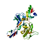
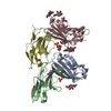
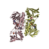
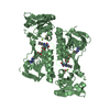
| Title | Structure of a Dimeric Lac Repressor with C-terminal Deletion and K84L Substitution | ||||||
 Components Components | Lactose Operon Repressor | ||||||
 Keywords Keywords | TRANSCRIPTION / Gene Regulation / Protein Stability / Protein DNA-binding | ||||||
| Function / homology |  Function and homology information Function and homology informationDNA-binding transcription repressor activity / cis-regulatory region sequence-specific DNA binding / transcription cis-regulatory region binding / DNA-binding transcription factor activity / negative regulation of DNA-templated transcription / regulation of DNA-templated transcription / DNA-templated transcription / identical protein binding / cytosol Similarity search - Function | ||||||
| Biological species |  | ||||||
| Method |  X-RAY DIFFRACTION / SYNCHROTRON / MOLECULAR REPLACEMENT / Resolution: 1.7 Å X-RAY DIFFRACTION / SYNCHROTRON / MOLECULAR REPLACEMENT / Resolution: 1.7 Å | ||||||
 Authors Authors | Bell, C.E. / Barry, J. / Matthews, K.S. / Lewis, M. | ||||||
 Citation Citation | Journal: J.Mol.Biol. / Year: 2001 Title: Structure of a variant of lac repressor with increased thermostability and decreased affinity for operator. Authors: Bell, C.E. / Barry, J. / Matthews, K.S. / Lewis, M. | ||||||
| History |
|
- Structure visualization
| Structure viewer | Molecule: MolmilJmol/JSmol |
|---|
- Downloads & links
-Download
| PDBx/mmCIF format | 1jye.cif.gz | 70 KB | Display | PDBx/mmCIF format |
|---|---|---|---|---|
| PDB format | pdb1jye.ent.gz | 51 KB | Display | PDB format |
| PDBx/mmJSON format | 1jye.json.gz | Tree view | PDBx/mmJSON format | |
| Others |  Other downloads Other downloads |
-Validation report
| Arichive directory | https://data.pdbj.org/pub/pdb/validation_reports/jy/1jyeftp://data.pdbj.org/pub/pdb/validation_reports/jy/1jye | HTTPS FTP |
|---|
-Related structure data


-Links
 PDBj
PDBj


- Assembly
| Deposited unit | 
| |||||||||||||||||||||
|---|---|---|---|---|---|---|---|---|---|---|---|---|---|---|---|---|---|---|---|---|---|---|
| 1 | 
| |||||||||||||||||||||
| Unit cell |
| |||||||||||||||||||||
| Components on special symmetry positions |
|
-Components
| #1: Protein | Mass: 37426.691 Da / Num. of mol.: 1 / Fragment: C-TERMINAL DELETION MUTANT / Mutation: YES Source method: isolated from a genetically manipulated source Source: (gene. exp.) |
|---|---|
| #2: Chemical | ChemComp-GOL /   Mass: 92.094 Da / Num. of mol.: 1 / Source method: obtained synthetically / Formula: C3H8O3 Mass: 92.094 Da / Num. of mol.: 1 / Source method: obtained synthetically / Formula: C3H8O3 |
| #3: Water | ChemComp-HOH /  Mass: 18.015 Da / Num. of mol.: 235 / Source method: isolated from a natural source / Formula: H2O Mass: 18.015 Da / Num. of mol.: 235 / Source method: isolated from a natural source / Formula: H2O |
| Sequence details | THE DIFFERENCE AT POSITION 109 IS A DIFFERENCE IN THE SEQUENCE REPORTED BY FARABAUGH, P.J. (1978) ...THE DIFFERENCE |
-Experimental details
-Experiment
| Experiment | Method: X-RAY DIFFRACTION / Number of used crystals: 1 |
|---|
- Sample preparation
| Crystal | Density Matthews: 2.83 Å3/Da / Density % sol: 56.57 % | |||||||||||||||
|---|---|---|---|---|---|---|---|---|---|---|---|---|---|---|---|---|
| Crystal grow | Temperature: 295 K / Method: vapor diffusion, hanging drop / pH: 5.6 Details: Sodium Acetate, Sodium Citrate, pH 5.6, VAPOR DIFFUSION, HANGING DROP, temperature 295K | |||||||||||||||
| Crystal | *PLUS Density % sol: 50 % | |||||||||||||||
| Crystal grow | *PLUS | |||||||||||||||
| Components of the solutions | *PLUS
|
-Data collection
| Diffraction | Mean temperature: 100 K |
|---|---|
| Diffraction source | Source: SYNCHROTRON / Site: NSLS  / Beamline: X25 / Wavelength: 1.1 / Beamline: X25 / Wavelength: 1.1 |
| Detector | Type: BRANDEIS - B4 / Detector: CCD |
| Radiation | Protocol: SINGLE WAVELENGTH / Monochromatic (M) / Laue (L): M / Scattering type: x-ray |
| Radiation wavelength | Wavelength: 1.1 Å / Relative weight: 1 |
| Reflection | Resolution: 1.7→17 Å / Num. obs: 44403 / % possible obs: 93.1 % / Observed criterion σ(F): 0 / Observed criterion σ(I): -2 / Redundancy: 5.8 % / Biso Wilson estimate: 23.4 Å2 / Rmerge(I) obs: 0.063 / Net I/σ(I): 12.5 |
| Reflection shell | Highest resolution: 1.7 Å / Rmerge(I) obs: 0.089 / Mean I/σ(I) obs: 7.2 / % possible all: 72.7 |
| Reflection | *PLUS Num. measured all: 258845 |
| Reflection shell | *PLUS % possible obs: 72.7 % |
- Processing
| Software |
| ||||||||||||||||||||||||||||||||||||||||
|---|---|---|---|---|---|---|---|---|---|---|---|---|---|---|---|---|---|---|---|---|---|---|---|---|---|---|---|---|---|---|---|---|---|---|---|---|---|---|---|---|---|
| Refinement | Method to determine structure: MOLECULAR REPLACEMENT Starting model: lac repressor Resolution: 1.7→9.95 Å / Rfactor Rfree error: 0.004 / Data cutoff high absF: 1304025.4 / Data cutoff high rms absF: 1304025.4 / Data cutoff low absF: 0 / Isotropic thermal model: RESTRAINED / Cross valid method: THROUGHOUT / σ(F): 0
| ||||||||||||||||||||||||||||||||||||||||
| Solvent computation | Solvent model: FLAT MODEL / Bsol: 66.1617 Å2 / ksol: 0.520559 e/Å3 | ||||||||||||||||||||||||||||||||||||||||
| Displacement parameters | Biso mean: 22 Å2
| ||||||||||||||||||||||||||||||||||||||||
| Refine analyze |
| ||||||||||||||||||||||||||||||||||||||||
| Refinement step | Cycle: LAST / Resolution: 1.7→9.95 Å
| ||||||||||||||||||||||||||||||||||||||||
| Refine LS restraints |
| ||||||||||||||||||||||||||||||||||||||||
| LS refinement shell | Resolution: 1.7→1.81 Å / Rfactor Rfree error: 0.012 / Total num. of bins used: 6
| ||||||||||||||||||||||||||||||||||||||||
| Xplor file |
| ||||||||||||||||||||||||||||||||||||||||
| Software | *PLUS Name: CNS / Version: 1 / Classification: refinement | ||||||||||||||||||||||||||||||||||||||||
| Refinement | *PLUS σ(F): 0 / % reflection Rfree: 10 % / Rfactor obs: 0.223 | ||||||||||||||||||||||||||||||||||||||||
| Solvent computation | *PLUS | ||||||||||||||||||||||||||||||||||||||||
| Displacement parameters | *PLUS Biso mean: 22 Å2 | ||||||||||||||||||||||||||||||||||||||||
| Refine LS restraints | *PLUS
| ||||||||||||||||||||||||||||||||||||||||
| LS refinement shell | *PLUS Rfactor Rfree: 0.284 / % reflection Rfree: 10.1 % / Rfactor Rwork: 0.284 |
