 Movie
Movie Controller


+ Open data

- Basic information
| Entry | Database: PDB / ID: 1jwr | ||||||
|---|---|---|---|---|---|---|---|
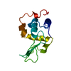
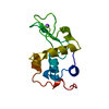
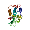
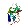
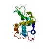
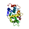
| Title | Crystal structure of human lysozyme at 100 K | ||||||
 Components Components | lysozyme | ||||||
 Keywords Keywords | HYDROLASE / hydration structure | ||||||
| Function / homology |  Function and homology information Function and homology informationantimicrobial humoral response / Antimicrobial peptides / specific granule lumen / azurophil granule lumen / lysozyme / lysozyme activity / tertiary granule lumen / defense response to Gram-negative bacterium / killing of cells of another organism / defense response to Gram-positive bacterium ...antimicrobial humoral response / Antimicrobial peptides / specific granule lumen / azurophil granule lumen / lysozyme / lysozyme activity / tertiary granule lumen / defense response to Gram-negative bacterium / killing of cells of another organism / defense response to Gram-positive bacterium / defense response to bacterium / Amyloid fiber formation / inflammatory response / Neutrophil degranulation / extracellular space / extracellular exosome / extracellular region / identical protein binding Similarity search - Function | ||||||
| Biological species |  Homo sapiens (human) Homo sapiens (human) | ||||||
| Method |  X-RAY DIFFRACTION / MOLECULAR REPLACEMENT / Resolution: 1.4 Å X-RAY DIFFRACTION / MOLECULAR REPLACEMENT / Resolution: 1.4 Å | ||||||
 Authors Authors | Higo, J. / Nakasako, M. | ||||||
 Citation Citation | Journal: J.Comput.Chem. / Year: 2002 Title: Hydration structure of human lysozyme investigated by molecular dynamics simulation and cryogenic X-ray crystal structure analyses: on the correlation between crystal water sites, solvent ...Title: Hydration structure of human lysozyme investigated by molecular dynamics simulation and cryogenic X-ray crystal structure analyses: on the correlation between crystal water sites, solvent density, and solvent dipole Authors: Higo, J. / Nakasako, M. | ||||||
| History |
|
- Structure visualization
| Structure viewer | Molecule: MolmilJmol/JSmol |
|---|
- Downloads & links
-Download
| PDBx/mmCIF format | 1jwr.cif.gz | 44.4 KB | Display | PDBx/mmCIF format |
|---|---|---|---|---|
| PDB format | pdb1jwr.ent.gz | 30.4 KB | Display | PDB format |
| PDBx/mmJSON format | 1jwr.json.gz | Tree view | PDBx/mmJSON format | |
| Others |  Other downloads Other downloads |
-Validation report
| Summary document | 1jwr_validation.pdf.gz | 350 KB | Display | wwPDB validaton report |
|---|---|---|---|---|
| Full document | 1jwr_full_validation.pdf.gz | 351.5 KB | Display | |
| Data in XML | 1jwr_validation.xml.gz | 3.7 KB | Display | |
| Data in CIF | 1jwr_validation.cif.gz | 7 KB | Display | |
| Arichive directory | https://data.pdbj.org/pub/pdb/validation_reports/jw/1jwrftp://data.pdbj.org/pub/pdb/validation_reports/jw/1jwr | HTTPS FTP |
-Related structure data
| Related structure data |  1rexS S: Starting model for refinement |
|---|---|
| Similar structure data |


-Links
 PDBj
PDBj

- Assembly
| Deposited unit | 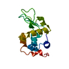
| ||||||||
|---|---|---|---|---|---|---|---|---|---|
| 1 |
| ||||||||
| Unit cell |
|
-Components
| #1: Protein | Mass: 14720.693 Da / Num. of mol.: 1 Source method: isolated from a genetically manipulated source Source: (gene. exp.) Homo sapiens (human) / Production host:  |
|---|---|
| #2: Water | ChemComp-HOH /  Mass: 18.015 Da / Num. of mol.: 307 / Source method: isolated from a natural source / Formula: H2O Mass: 18.015 Da / Num. of mol.: 307 / Source method: isolated from a natural source / Formula: H2O |
| Has protein modification | Y |
-Experimental details
-Experiment
| Experiment | Method: X-RAY DIFFRACTION / Number of used crystals: 1 |
|---|
- Sample preparation
| Crystal | Density Matthews: 1.93 Å3/Da / Density % sol: 36.24 % | ||||||||||||||||||||||||
|---|---|---|---|---|---|---|---|---|---|---|---|---|---|---|---|---|---|---|---|---|---|---|---|---|---|
| Crystal grow | Temperature: 293 K / Method: vapor diffusion, hanging drop / pH: 4.5 Details: NaCl, sodium acetate, pH 4.5, VAPOR DIFFUSION, HANGING DROP, temperature 293K | ||||||||||||||||||||||||
| Crystal grow | *PLUS Temperature: 10 ℃ / Details: Takano, K., (1995) J. Mol. Biol., 254, 62. | ||||||||||||||||||||||||
| Components of the solutions | *PLUS
|
-Data collection
| Diffraction | Mean temperature: 100 K |
|---|---|
| Diffraction source | Source: ROTATING ANODE / Type: RIGAKU ULTRAX 18H / Wavelength: 1.5418 Å |
| Detector | Type: RIGAKU RAXIS IV / Detector: IMAGE PLATE / Date: Jan 1, 1998 / Details: double-focusing Pt-coated mirrors |
| Radiation | Monochromator: Ni filter / Protocol: SINGLE WAVELENGTH / Monochromatic (M) / Laue (L): M / Scattering type: x-ray |
| Radiation wavelength | Wavelength: 1.5418 Å / Relative weight: 1 |
| Reflection | Resolution: 1.4→50 Å / Num. all: 184173 / Num. obs: 22814 / % possible obs: 98.4 % / Observed criterion σ(I): 1 / Redundancy: 8.07 % / Rmerge(I) obs: 0.048 / Net I/σ(I): 44.6 |
| Reflection shell | Resolution: 1.4→1.42 Å / Redundancy: 7.95 % / Rmerge(I) obs: 0.115 / Mean I/σ(I) obs: 11.3 / Num. unique all: 811 / % possible all: 94.9 |
| Reflection | *PLUS Num. measured all: 184173 |
| Reflection shell | *PLUS % possible obs: 94.9 % |
- Processing
| Software |
| |||||||||||||||||||||||||
|---|---|---|---|---|---|---|---|---|---|---|---|---|---|---|---|---|---|---|---|---|---|---|---|---|---|---|
| Refinement | Method to determine structure: MOLECULAR REPLACEMENT Starting model: PDB entry 1rex Resolution: 1.4→8 Å / Isotropic thermal model: isotropic / Cross valid method: THROUGHOUT / σ(F): 2 / Stereochemistry target values: Engh & Huber
| |||||||||||||||||||||||||
| Refine analyze | Luzzati coordinate error obs: 0.016 Å | |||||||||||||||||||||||||
| Refinement step | Cycle: LAST / Resolution: 1.4→8 Å
| |||||||||||||||||||||||||
| Refine LS restraints |
| |||||||||||||||||||||||||
| Software | *PLUS Name: X-PLOR / Classification: refinement | |||||||||||||||||||||||||
| Refinement | *PLUS σ(F): 2 / % reflection Rfree: 11 % / Rfactor obs: 0.178 | |||||||||||||||||||||||||
| Solvent computation | *PLUS | |||||||||||||||||||||||||
| Displacement parameters | *PLUS | |||||||||||||||||||||||||
| Refine LS restraints | *PLUS
|
