 Movie
Movie Controller

[English] 日本語
 Yorodumi
- PDB-1jux: 1.6A Resolution Crystal Structures of the DNA Octamers d(IUATATAC... -
+ Open data

- Basic information
| Entry | Database: PDB / ID: 1jux | ||||||||||||||||||
|---|---|---|---|---|---|---|---|---|---|---|---|---|---|---|---|---|---|---|---|
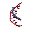
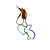
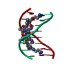
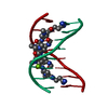
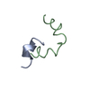
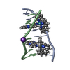
| Title | 1.6A Resolution Crystal Structures of the DNA Octamers d(IUATATAC) and d(ITITACAC):Binding of Two Distamycin Drugs Side-by-Side | ||||||||||||||||||
 Components Components | 5'-D(* Keywords KeywordsDNA / Octamer / Distamycin / Side-by-Side | Function / homology | DISTAMYCIN A / DNA |  Function and homology information Function and homology informationMethod |  X-RAY DIFFRACTION / MOLECULAR REPLACEMENT / Resolution: 1.6 Å X-RAY DIFFRACTION / MOLECULAR REPLACEMENT / Resolution: 1.6 Å  Authors AuthorsDeng, J. / Pan, B. / Sundaralingam, M. |  CitationJournal: Acta Crystallogr.,Sect.D / Year: 2003 CitationJournal: Acta Crystallogr.,Sect.D / Year: 2003Title: Structure of d(ITITACAC) complexed with distamycin at 1.6 A resolution. Authors: Deng, J. / Pan, B. / Sundaralingam, M. History |
|
- Structure visualization
| Structure viewer | Molecule: MolmilJmol/JSmol |
|---|
- Downloads & links
-Download
| PDBx/mmCIF format | 1jux.cif.gz | 14.1 KB | Display | PDBx/mmCIF format |
|---|---|---|---|---|
| PDB format | pdb1jux.ent.gz | 8.5 KB | Display | PDB format |
| PDBx/mmJSON format | 1jux.json.gz | Tree view | PDBx/mmJSON format | |
| Others |  Other downloads Other downloads |
-Validation report
| Arichive directory | https://data.pdbj.org/pub/pdb/validation_reports/ju/1juxftp://data.pdbj.org/pub/pdb/validation_reports/ju/1jux | HTTPS FTP |
|---|
-Related structure data
| Related structure data |  1juz |
|---|---|
| Similar structure data |


-Links
 PDBj
PDBj

- Assembly
| Deposited unit | 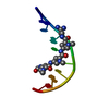
| ||||||||
|---|---|---|---|---|---|---|---|---|---|
| 1 | 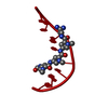
| ||||||||
| Unit cell |
| ||||||||
| Details | One strand in the assymetric unit. The duplex is generated by the 2 fold axis. |
-Components
| #1: DNA chain | Mass: 2396.589 Da / Num. of mol.: 1 / Source method: obtained synthetically / Details: Octamer DNA |
|---|---|
| #2: Chemical | ChemComp-DMY /   Mass: 481.508 Da / Num. of mol.: 1 / Source method: obtained synthetically / Formula: C22H27N9O4 / Comment: antibiotic*YM Mass: 481.508 Da / Num. of mol.: 1 / Source method: obtained synthetically / Formula: C22H27N9O4 / Comment: antibiotic*YM |
| #3: Water | ChemComp-HOH /  Mass: 18.015 Da / Num. of mol.: 36 / Source method: isolated from a natural source / Formula: H2O Mass: 18.015 Da / Num. of mol.: 36 / Source method: isolated from a natural source / Formula: H2O |
-Experimental details
-Experiment
| Experiment | Method: X-RAY DIFFRACTION / Number of used crystals: 1 |
|---|
- Sample preparation
| Crystal | Density Matthews: 1.96 Å3/Da / Density % sol: 37.09 % | ||||||||||||||||||||||||||||||||||||
|---|---|---|---|---|---|---|---|---|---|---|---|---|---|---|---|---|---|---|---|---|---|---|---|---|---|---|---|---|---|---|---|---|---|---|---|---|---|
| Crystal grow | *PLUS pH: 7 / Method: vapor diffusion, hanging drop | ||||||||||||||||||||||||||||||||||||
| Components of the solutions | *PLUS
|
-Data collection
| Diffraction | Mean temperature: 298 K |
|---|---|
| Diffraction source | Source: ROTATING ANODE / Type: RIGAKU RU200 / Wavelength: 1.5418 Å |
| Detector | Type: RIGAKU RAXIS IIC / Detector: IMAGE PLATE / Date: Jun 20, 1997 |
| Radiation | Monochromator: graphite / Protocol: SINGLE WAVELENGTH / Monochromatic (M) / Laue (L): M / Scattering type: x-ray |
| Radiation wavelength | Wavelength: 1.5418 Å / Relative weight: 1 |
| Reflection | Resolution: 1.6→10 Å / Num. all: 2609 / Num. obs: 2468 / Observed criterion σ(F): 2 |
| Reflection | *PLUS Highest resolution: 1.6 Å / Lowest resolution: 10 Å / Num. obs: 2737 / % possible obs: 92 % / Rmerge(I) obs: 0.046 |
| Reflection shell | *PLUS Highest resolution: 1.6 Å / Lowest resolution: 1.7 Å / % possible obs: 88.8 % / Num. unique obs: 381 / Rmerge(I) obs: 0.162 |
- Processing
| Software |
| |||||||||||||||||||||||||
|---|---|---|---|---|---|---|---|---|---|---|---|---|---|---|---|---|---|---|---|---|---|---|---|---|---|---|
| Refinement | Method to determine structure: MOLECULAR REPLACEMENT / Resolution: 1.6→10 Å / σ(F): 2
| |||||||||||||||||||||||||
| Refinement step | Cycle: LAST / Resolution: 1.6→10 Å
| |||||||||||||||||||||||||
| Refinement | *PLUS Lowest resolution: 10 Å / Num. reflection obs: 2520 / Rfactor Rfree: 0.207 / Rfactor Rwork: 0.17 | |||||||||||||||||||||||||
| Solvent computation | *PLUS | |||||||||||||||||||||||||
| Displacement parameters | *PLUS | |||||||||||||||||||||||||
| Refine LS restraints | *PLUS
|
