 Movie
Movie Controller

[English] 日本語
 Yorodumi
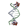
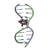
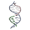
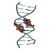
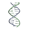
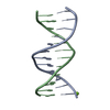
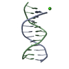
- PDB-1juu: NMR Structure of a Parallel Stranded DNA Duplex at Atomic Resolution -
+ Open data

- Basic information
| Entry | Database: PDB / ID: 1juu | ||||||
|---|---|---|---|---|---|---|---|
| Title | NMR Structure of a Parallel Stranded DNA Duplex at Atomic Resolution | ||||||
 Components Components |
| ||||||
 Keywords Keywords | DNA / Parallel stranded / DNA duplex | ||||||
| Function / homology | DNA / DNA (> 10) Function and homology information Function and homology information | ||||||
| Method | SOLUTION NMR / simulated annealing, molecular dynamics | ||||||
 Authors Authors | Parvathy, V.R. / Bhaumik, S.R. / Chary, K.V.R. / Govil, G. / Liu, K. / Howard, F.B. / Miles, H.T. | ||||||
 Citation Citation | Journal: Nucleic Acids Res. / Year: 2002 Title: NMR structure of a parallel-stranded DNA duplex at atomic resolution. Authors: Parvathy, V.R. / Bhaumik, S.R. / Chary, K.V. / Govil, G. / Liu, K. / Howard, F.B. / Miles, H.T. | ||||||
| History |
|
- Structure visualization
| Structure viewer | Molecule: MolmilJmol/JSmol |
|---|
- Downloads & links
-Download
| PDBx/mmCIF format | 1juu.cif.gz | 299.2 KB | Display | PDBx/mmCIF format |
|---|---|---|---|---|
| PDB format | pdb1juu.ent.gz | 245 KB | Display | PDB format |
| PDBx/mmJSON format | 1juu.json.gz | Tree view | PDBx/mmJSON format | |
| Others |  Other downloads Other downloads |
-Validation report
| Arichive directory | https://data.pdbj.org/pub/pdb/validation_reports/ju/1juuftp://data.pdbj.org/pub/pdb/validation_reports/ju/1juu | HTTPS FTP |
|---|
-Related structure data
| Similar structure data |
|---|


-Links
 PDBj
PDBj


- Assembly
| Deposited unit | 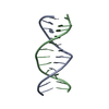
| |||||||||
|---|---|---|---|---|---|---|---|---|---|---|
| 1 |
| |||||||||
| NMR ensembles |
|
-Components
| #1: DNA chain | Mass: 3581.368 Da / Num. of mol.: 1 / Source method: obtained synthetically Details: The DNA duplex was Synthesized with an Applied Biosystems Model 380B DNA synthesizer using solid state phosphoramidate chemistry |
|---|---|
| #2: DNA chain | Mass: 3581.368 Da / Num. of mol.: 1 / Source method: obtained synthetically |
-Experimental details
-Experiment
| Experiment | Method: SOLUTION NMR |
|---|---|
| NMR experiment | Type: COSY, NOESY, TOCSY |
| NMR details | Text: Structure was determined using 2D homonuclear experiments. |
- Sample preparation
| Details | Contents: 5 mM concentration of each dodecamer strand, 0.05 M deuterated sodium acetate buffer and 0.1 M NaCl Solvent system: 90% H2O/10% D2O |
|---|---|
| Sample conditions | Ionic strength: 100mM NaCl / pH: 5.5 / Pressure: ambient / Temperature: 303 K |
| Crystal grow | *PLUS Method: other / Details: NMR |
-NMR measurement
| Radiation | Protocol: SINGLE WAVELENGTH / Monochromatic (M) / Laue (L): M |
|---|---|
| Radiation wavelength | Relative weight: 1 |
| NMR spectrometer | Type: Varian UNITY / Manufacturer: Varian / Model: UNITY / Field strength: 600 MHz |
- Processing
| NMR software |
| ||||||||||||
|---|---|---|---|---|---|---|---|---|---|---|---|---|---|
| Refinement | Method: simulated annealing, molecular dynamics / Software ordinal: 1 Details: Structure was calculated using 78 nOe constraints, 28 hydrogen bond constraints, and 162 dihedral angle constraints. | ||||||||||||
| NMR representative | Selection criteria: fewest violations,lowest energy | ||||||||||||
| NMR ensemble | Conformer selection criteria: structures with the lowest energy Conformers calculated total number: 200 / Conformers submitted total number: 20 |
