 Movie
Movie Controller

[English] 日本語
 Yorodumi
- PDB-1jut: E. coli Thymidylate Synthase Bound to dUMP and LY338529, A Pyrrol... -
+ Open data

- Basic information
| Entry | Database: PDB / ID: 1jut | ||||||
|---|---|---|---|---|---|---|---|
| Title | E. coli Thymidylate Synthase Bound to dUMP and LY338529, A Pyrrolo(2,3-d)pyrimidine-based Antifolate | ||||||
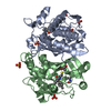
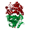
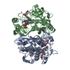
 Components Components | THYMIDYLATE SYNTHASE | ||||||
 Keywords Keywords | TRANSFERASE / antifolate / dTMP synthesis / cancer / drug resistance | ||||||
| Function / homology |  Function and homology information Function and homology informationthymidylate synthase / thymidylate synthase activity / dTTP biosynthetic process / dTMP biosynthetic process / response to radiation / regulation of translation / methylation / magnesium ion binding / protein homodimerization activity / RNA binding / cytosol Similarity search - Function | ||||||
| Biological species |  | ||||||
| Method |  X-RAY DIFFRACTION / FOURIER SYNTHESIS / Resolution: 2.7 Å X-RAY DIFFRACTION / FOURIER SYNTHESIS / Resolution: 2.7 Å | ||||||
 Authors Authors | Sayre, P.H. / Finer-Moore, J.S. / Fritz, T.A. / Biermann, D. / Gates, S.B. / MacKellar, W.C. / Patel, V.F. / Stroud, R.M. | ||||||
 Citation Citation | Journal: J.Mol.Biol. / Year: 2001 Title: Multi-targeted antifolates aimed at avoiding drug resistance form covalent closed inhibitory complexes with human and Escherichia coli thymidylate synthases. Authors: Sayre, P.H. / Finer-Moore, J.S. / Fritz, T.A. / Biermann, D. / Gates, S.B. / MacKellar, W.C. / Patel, V.F. / Stroud, R.M. | ||||||
| History |
|
- Structure visualization
| Structure viewer | Molecule: MolmilJmol/JSmol |
|---|
- Downloads & links
-Download
| PDBx/mmCIF format | 1jut.cif.gz | 120.8 KB | Display | PDBx/mmCIF format |
|---|---|---|---|---|
| PDB format | pdb1jut.ent.gz | 95.1 KB | Display | PDB format |
| PDBx/mmJSON format | 1jut.json.gz | Tree view | PDBx/mmJSON format | |
| Others |  Other downloads Other downloads |
-Validation report
| Arichive directory | https://data.pdbj.org/pub/pdb/validation_reports/ju/1jutftp://data.pdbj.org/pub/pdb/validation_reports/ju/1jut | HTTPS FTP |
|---|
-Related structure data
| Related structure data | 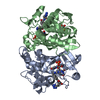 1jtqC 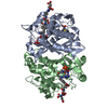 1jtuC 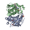 1ju6C 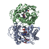 1jujC  2kceS C: citing same article ( S: Starting model for refinement |
|---|---|
| Similar structure data |


-Links
 PDBj
PDBj
- Assembly
| Deposited unit | 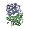
| ||||||||
|---|---|---|---|---|---|---|---|---|---|
| 1 |
| ||||||||
| Unit cell |
|
-Components
| #1: Protein | Mass: 30559.666 Da / Num. of mol.: 2 Source method: isolated from a genetically manipulated source Source: (gene. exp.) #2: Chemical |   Mass: 308.182 Da / Num. of mol.: 2 / Source method: obtained synthetically / Formula: C9H13N2O8P Mass: 308.182 Da / Num. of mol.: 2 / Source method: obtained synthetically / Formula: C9H13N2O8P#3: Chemical | 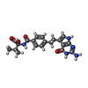  Mass: 397.428 Da / Num. of mol.: 2 / Source method: obtained synthetically / Formula: C20H23N5O4 Mass: 397.428 Da / Num. of mol.: 2 / Source method: obtained synthetically / Formula: C20H23N5O4#4: Water | ChemComp-HOH / |  Mass: 18.015 Da / Num. of mol.: 82 / Source method: isolated from a natural source / Formula: H2O Mass: 18.015 Da / Num. of mol.: 82 / Source method: isolated from a natural source / Formula: H2OHas protein modification | Y | |
|---|
-Experimental details
-Experiment
| Experiment | Method: X-RAY DIFFRACTION / Number of used crystals: 1 |
|---|
- Sample preparation
| Crystal | Density Matthews: 2.51 Å3/Da / Density % sol: 50.92 % | ||||||||||||||||||||||||||||||||||||||||||
|---|---|---|---|---|---|---|---|---|---|---|---|---|---|---|---|---|---|---|---|---|---|---|---|---|---|---|---|---|---|---|---|---|---|---|---|---|---|---|---|---|---|---|---|
| Crystal grow | Temperature: 296 K / Method: vapor diffusion, hanging drop / pH: 8 Details: KPO4, MgCl2, DTT, EDTA, ammonium sulphate, pH 8, VAPOR DIFFUSION, HANGING DROP, temperature 296K | ||||||||||||||||||||||||||||||||||||||||||
| Crystal grow | *PLUS pH: 6.5 | ||||||||||||||||||||||||||||||||||||||||||
| Components of the solutions | *PLUS
|
-Data collection
| Diffraction | Mean temperature: 296 K |
|---|---|
| Diffraction source | Source: ROTATING ANODE / Type: RIGAKU / Wavelength: 1.54178 Å |
| Detector | Type: RIGAKU RAXIS IV / Detector: IMAGE PLATE / Date: Apr 28, 1998 |
| Radiation | Monochromator: graphite / Protocol: SINGLE WAVELENGTH / Monochromatic (M) / Laue (L): M / Scattering type: x-ray |
| Radiation wavelength | Wavelength: 1.54178 Å / Relative weight: 1 |
| Reflection | Resolution: 2.7→30 Å / Num. all: 16866 / Num. obs: 16866 / % possible obs: 100 % / Observed criterion σ(F): 0 / Observed criterion σ(I): -3 / Redundancy: 5.8 % / Biso Wilson estimate: 44.9 Å2 / Rmerge(I) obs: 0.163 / Net I/σ(I): 8 |
| Reflection shell | Resolution: 2.7→2.8 Å / Rmerge(I) obs: 0.6 / Mean I/σ(I) obs: 1.9 / % possible all: 99 |
| Reflection | *PLUS Lowest resolution: 30 Å / % possible obs: 100 % / Num. measured all: 97881 |
| Reflection shell | *PLUS % possible obs: 99 % / Rmerge(I) obs: 0.599 |
- Processing
| Software |
| |||||||||||||||||||||||||
|---|---|---|---|---|---|---|---|---|---|---|---|---|---|---|---|---|---|---|---|---|---|---|---|---|---|---|
| Refinement | Method to determine structure: FOURIER SYNTHESIS Starting model: PDB entry 2KCE Resolution: 2.7→29.59 Å / Rfactor Rfree error: 0.007 / Data cutoff high absF: 1797097.09 / Data cutoff low absF: 0 / Isotropic thermal model: RESTRAINED / Cross valid method: THROUGHOUT / σ(F): 0 / Stereochemistry target values: Engh and Huber
| |||||||||||||||||||||||||
| Solvent computation | Solvent model: FLAT MODEL / Bsol: 28.6405 Å2 / ksol: 0.365014 e/Å3 | |||||||||||||||||||||||||
| Displacement parameters | Biso mean: 30.8 Å2
| |||||||||||||||||||||||||
| Refine analyze |
| |||||||||||||||||||||||||
| Refinement step | Cycle: LAST / Resolution: 2.7→29.59 Å
| |||||||||||||||||||||||||
| Refine LS restraints |
| |||||||||||||||||||||||||
| LS refinement shell | Resolution: 2.7→2.87 Å / Rfactor Rfree error: 0.027 / Total num. of bins used: 6
| |||||||||||||||||||||||||
| Xplor file |
| |||||||||||||||||||||||||
| Software | *PLUS Name: CNS / Version: 1 / Classification: refinement | |||||||||||||||||||||||||
| Refinement | *PLUS σ(F): 0 / % reflection Rfree: 6.9 % / Rfactor obs: 0.22 / Rfactor Rfree: 0.25 | |||||||||||||||||||||||||
| Solvent computation | *PLUS | |||||||||||||||||||||||||
| Displacement parameters | *PLUS Biso mean: 30.8 Å2 | |||||||||||||||||||||||||
| Refine LS restraints | *PLUS
| |||||||||||||||||||||||||
| LS refinement shell | *PLUS Rfactor Rfree: 0.359 / % reflection Rfree: 6.6 % / Rfactor Rwork: 0.297 |
