 Movie
Movie Controller

[English] 日本語
 Yorodumi
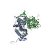
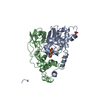
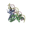
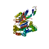
- PDB-1juo: Crystal Structure of Calcium-free Human Sorcin: A Member of the P... -
+ Open data

- Basic information
| Entry | Database: PDB / ID: 1juo | ||||||
|---|---|---|---|---|---|---|---|
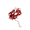
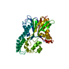
| Title | Crystal Structure of Calcium-free Human Sorcin: A Member of the Penta-EF-Hand Protein Family | ||||||
 Components Components | sorcin | ||||||
 Keywords Keywords | METAL TRANSPORT / calcium-binding proteins / penta-ef-hand / PEF | ||||||
| Function / homology |  Function and homology information Function and homology informationregulation of relaxation of muscle / regulation of cell communication by electrical coupling / negative regulation of cardiac muscle contraction / regulation of insulin secretion involved in cellular response to glucose stimulus / regulation of cardiac muscle cell contraction / Sodium/Calcium exchangers / negative regulation of heart rate / Reduction of cytosolic Ca++ levels / regulation of calcium ion transport / regulation of cell communication by electrical coupling involved in cardiac conduction ...regulation of relaxation of muscle / regulation of cell communication by electrical coupling / negative regulation of cardiac muscle contraction / regulation of insulin secretion involved in cellular response to glucose stimulus / regulation of cardiac muscle cell contraction / Sodium/Calcium exchangers / negative regulation of heart rate / Reduction of cytosolic Ca++ levels / regulation of calcium ion transport / regulation of cell communication by electrical coupling involved in cardiac conduction / Ion transport by P-type ATPases / transcription regulator inhibitor activity / regulation of release of sequestered calcium ion into cytosol by sarcoplasmic reticulum / Ion homeostasis / sarcoplasmic reticulum membrane / T-tubule / positive regulation of release of sequestered calcium ion into cytosol / sarcoplasmic reticulum / calcium channel regulator activity / protein sequestering activity / Stimuli-sensing channels / Z disc / calcium ion transport / protease binding / DNA-binding transcription factor binding / transmembrane transporter binding / protein heterodimerization activity / signaling receptor binding / calcium ion binding / endoplasmic reticulum membrane / signal transduction / extracellular exosome / nucleoplasm / membrane / identical protein binding / cytoplasm / cytosol Similarity search - Function | ||||||
| Biological species |  Homo sapiens (human) Homo sapiens (human) | ||||||
| Method |  X-RAY DIFFRACTION / MIR / Resolution: 2.2 Å X-RAY DIFFRACTION / MIR / Resolution: 2.2 Å | ||||||
 Authors Authors | Xie, X. | ||||||
 Citation Citation | Journal: Protein Sci. / Year: 2001 Title: Crystal structure of calcium-free human sorcin: a member of the penta-EF-hand protein family. Authors: Xie, X. / Dwyer, M.D. / Swenson, L. / Parker, M.H. / Botfield, M.C. | ||||||
| History |
|
- Structure visualization
| Structure viewer | Molecule: MolmilJmol/JSmol |
|---|
- Downloads & links
-Download
| PDBx/mmCIF format | 1juo.cif.gz | 83.8 KB | Display | PDBx/mmCIF format |
|---|---|---|---|---|
| PDB format | pdb1juo.ent.gz | 64 KB | Display | PDB format |
| PDBx/mmJSON format | 1juo.json.gz | Tree view | PDBx/mmJSON format | |
| Others |  Other downloads Other downloads |
-Validation report
| Arichive directory | https://data.pdbj.org/pub/pdb/validation_reports/ju/1juoftp://data.pdbj.org/pub/pdb/validation_reports/ju/1juo | HTTPS FTP |
|---|
-Related structure data
| Similar structure data |
|---|


-Links
 PDBj
PDBj


- Assembly
| Deposited unit | 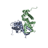
| ||||||||
|---|---|---|---|---|---|---|---|---|---|
| 1 |
| ||||||||
| Unit cell |
|
-Components
| #1: Protein | Mass: 21695.336 Da / Num. of mol.: 2 Source method: isolated from a genetically manipulated source Source: (gene. exp.) Homo sapiens (human) / Production host:  #2: Water | ChemComp-HOH / |  Mass: 18.015 Da / Num. of mol.: 285 / Source method: isolated from a natural source / Formula: H2O Mass: 18.015 Da / Num. of mol.: 285 / Source method: isolated from a natural source / Formula: H2O |
|---|
-Experimental details
-Experiment
| Experiment | Method: X-RAY DIFFRACTION / Number of used crystals: 1 |
|---|
- Sample preparation
| Crystal | Density Matthews: 3.44 Å3/Da / Density % sol: 64.26 % | |||||||||||||||||||||||||||||||||||||||||||||||||
|---|---|---|---|---|---|---|---|---|---|---|---|---|---|---|---|---|---|---|---|---|---|---|---|---|---|---|---|---|---|---|---|---|---|---|---|---|---|---|---|---|---|---|---|---|---|---|---|---|---|---|
| Crystal grow | Temperature: 298 K / Method: vapor diffusion, hanging drop / pH: 7 Details: sodium chloride, HEPES, EGTA, pH 7.0, VAPOR DIFFUSION, HANGING DROP, temperature 298.0K | |||||||||||||||||||||||||||||||||||||||||||||||||
| Crystal | *PLUS Density % sol: 64 % | |||||||||||||||||||||||||||||||||||||||||||||||||
| Crystal grow | *PLUS | |||||||||||||||||||||||||||||||||||||||||||||||||
| Components of the solutions | *PLUS
|
-Data collection
| Diffraction | Mean temperature: 100 K |
|---|---|
| Diffraction source | Source: ROTATING ANODE / Type: RIGAKU RU200 / Wavelength: 1.5418 Å |
| Detector | Type: RIGAKU RAXIS IV / Detector: IMAGE PLATE / Date: Nov 30, 1999 / Details: mirrors |
| Radiation | Monochromator: Confocal Multilayer (Rigaku/MSC) / Protocol: SINGLE WAVELENGTH / Monochromatic (M) / Laue (L): M / Scattering type: x-ray |
| Radiation wavelength | Wavelength: 1.5418 Å / Relative weight: 1 |
| Reflection | Resolution: 2.2→50 Å / Num. all: 167295 / Num. obs: 28472 / % possible obs: 90.7 % / Observed criterion σ(I): -1 / Biso Wilson estimate: 32.1 Å2 / Rmerge(I) obs: 0.064 |
| Reflection shell | Resolution: 2.2→2.26 Å / Rmerge(I) obs: 0.491 / Mean I/σ(I) obs: 2.1 / Num. unique all: 1929 / % possible all: 75.3 |
| Reflection | *PLUS Num. measured all: 167295 |
- Processing
| Software |
| |||||||||||||||||||||||||
|---|---|---|---|---|---|---|---|---|---|---|---|---|---|---|---|---|---|---|---|---|---|---|---|---|---|---|
| Refinement | Method to determine structure: MIR / Resolution: 2.2→37.56 Å / Rfactor Rfree error: 0.005 / Data cutoff high absF: 368354.7 / Data cutoff low absF: 0 / Isotropic thermal model: RESTRAINED / Cross valid method: THROUGHOUT / σ(F): 0 / Stereochemistry target values: Engh & Huber Details: RESOLUTION-DEPENDENT WEIGHTING SCHEME USED. BULK SOLVENT MODEL USED.
| |||||||||||||||||||||||||
| Solvent computation | Solvent model: flat model / Bsol: 41.8194 Å2 / ksol: 0.294676 e/Å3 | |||||||||||||||||||||||||
| Displacement parameters | Biso mean: 47.1 Å2
| |||||||||||||||||||||||||
| Refine analyze |
| |||||||||||||||||||||||||
| Refinement step | Cycle: LAST / Resolution: 2.2→37.56 Å
| |||||||||||||||||||||||||
| Refine LS restraints |
| |||||||||||||||||||||||||
| LS refinement shell | Resolution: 2.2→2.34 Å / Rfactor Rfree error: 0.018 / Total num. of bins used: 6
| |||||||||||||||||||||||||
| Xplor file |
| |||||||||||||||||||||||||
| Software | *PLUS Name: CNX2000 / Classification: refinement | |||||||||||||||||||||||||
| Refinement | *PLUS σ(F): 0 / % reflection Rfree: 9.9 % / Rfactor obs: 0.212 / Rfactor Rfree: 0.256 | |||||||||||||||||||||||||
| Solvent computation | *PLUS | |||||||||||||||||||||||||
| Displacement parameters | *PLUS Biso mean: 47.1 Å2 | |||||||||||||||||||||||||
| Refine LS restraints | *PLUS
| |||||||||||||||||||||||||
| LS refinement shell | *PLUS Rfactor Rfree: 0.363 / % reflection Rfree: 10.3 % / Rfactor Rwork: 0.347 |
