 Movie
Movie Controller


+ Open data

- Basic information
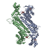
| Entry | Database: PDB / ID: 1jsw | ||||||
|---|---|---|---|---|---|---|---|
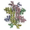
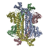
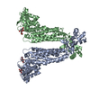
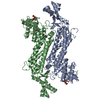
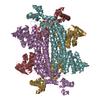
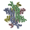
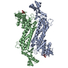
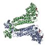
| Title | NATIVE L-ASPARTATE AMMONIA LYASE | ||||||
 Components Components | L-ASPARTATE AMMONIA-LYASE | ||||||
 Keywords Keywords | AMINO ACID AMMONIA-LYASE | ||||||
| Function / homology |  Function and homology information Function and homology informationaspartate ammonia-lyase / aspartate ammonia-lyase activity / L-aspartate catabolic process / nitrogen utilization / aspartate metabolic process / L-glutamate catabolic process / tricarboxylic acid cycle / protein homotetramerization / membrane / identical protein binding / cytosol Similarity search - Function | ||||||
| Biological species |  | ||||||
| Method |  X-RAY DIFFRACTION / Resolution: 2.7 Å X-RAY DIFFRACTION / Resolution: 2.7 Å | ||||||
 Authors Authors | Shi, W. / Dunbar, J. / Farber, G.K. | ||||||
 Citation Citation | Journal: Biochemistry / Year: 1997 Title: The structure of L-aspartate ammonia-lyase from Escherichia coli. Authors: Shi, W. / Dunbar, J. / Jayasekera, M.M. / Viola, R.E. / Farber, G.K. #1: Journal: J.Mol.Biol. / Year: 1993Title: Crystallization and Preliminary X-Ray Studies of L-Aspartase from Escherichia Coli Authors: Shi, W. / Kidd, R. / Giorgianni, F. / Schindler, J.F. / Viola, R.E. / Farber, G.K. | ||||||
| History |
|
- Structure visualization
| Structure viewer | Molecule: MolmilJmol/JSmol |
|---|
- Downloads & links
-Download
| PDBx/mmCIF format | 1jsw.cif.gz | 308.9 KB | Display | PDBx/mmCIF format |
|---|---|---|---|---|
| PDB format | pdb1jsw.ent.gz | 256.2 KB | Display | PDB format |
| PDBx/mmJSON format | 1jsw.json.gz | Tree view | PDBx/mmJSON format | |
| Others |  Other downloads Other downloads |
-Validation report
| Arichive directory | https://data.pdbj.org/pub/pdb/validation_reports/js/1jswftp://data.pdbj.org/pub/pdb/validation_reports/js/1jsw | HTTPS FTP |
|---|
-Related structure data
| Similar structure data |
|---|

-Links
 PDBj
PDBj


- Assembly
| Deposited unit | 
| ||||||||
|---|---|---|---|---|---|---|---|---|---|
| 1 |
| ||||||||
| Unit cell |
|
-Components
| #1: Protein | Mass: 52406.883 Da / Num. of mol.: 4 / Source method: isolated from a natural source / Source: (natural) #2: Sugar |   Type: D-saccharide, beta linking / Mass: 180.156 Da / Num. of mol.: 2 Type: D-saccharide, beta linking / Mass: 180.156 Da / Num. of mol.: 2Source method: isolated from a genetically manipulated source Formula: C6H12O6 #3: Chemical | ChemComp-ACT / |   Mass: 59.044 Da / Num. of mol.: 1 / Source method: obtained synthetically / Formula: C2H3O2 Mass: 59.044 Da / Num. of mol.: 1 / Source method: obtained synthetically / Formula: C2H3O2#4: Water | ChemComp-HOH / |  Mass: 18.015 Da / Num. of mol.: 69 / Source method: isolated from a natural source / Formula: H2O Mass: 18.015 Da / Num. of mol.: 69 / Source method: isolated from a natural source / Formula: H2O |
|---|
-Experimental details
-Experiment
| Experiment | Method: X-RAY DIFFRACTION / Number of used crystals: 1 |
|---|
- Sample preparation
| Crystal | Density Matthews: 2.77 Å3/Da / Density % sol: 56 % | |||||||||||||||||||||||||||||||||||
|---|---|---|---|---|---|---|---|---|---|---|---|---|---|---|---|---|---|---|---|---|---|---|---|---|---|---|---|---|---|---|---|---|---|---|---|---|
| Crystal grow | *PLUS Temperature: 21 ℃ / pH: 7.5 / Method: microdialysis | |||||||||||||||||||||||||||||||||||
| Components of the solutions | *PLUS
|
-Data collection
| Diffraction | Mean temperature: 95 K |
|---|---|
| Diffraction source | Wavelength: 1.5418 |
| Detector | Type: RIGAKU RAXIS II / Detector: IMAGE PLATE / Date: Apr 26, 1996 |
| Radiation | Monochromator: GRAPHITE(002) / Monochromatic (M) / Laue (L): M / Scattering type: x-ray |
| Radiation wavelength | Wavelength: 1.5418 Å / Relative weight: 1 |
| Reflection | Num. obs: 57964 / % possible obs: 80.3 % / Redundancy: 2.7 % / Rmerge(I) obs: 0.076 |
| Reflection | *PLUS Highest resolution: 2.8 Å / Num. measured all: 156675 |
- Processing
| Software |
| ||||||||||||||||||||||||||||||||||||||||||||||||||||||||||||
|---|---|---|---|---|---|---|---|---|---|---|---|---|---|---|---|---|---|---|---|---|---|---|---|---|---|---|---|---|---|---|---|---|---|---|---|---|---|---|---|---|---|---|---|---|---|---|---|---|---|---|---|---|---|---|---|---|---|---|---|---|---|
| Refinement | Resolution: 2.7→10 Å / σ(F): 2 Details: RESIDUE 32 WAS MODELED AS A VAL FOR MOST OF THE REFINEMENT CYCLES BECAUSE OF CONFLICTING SEQUENCES AT THIS POSITION. A SEQUENCING GEL CONFIRMED THAT THIS RESIDUE IS ACTUALLY GLU. THE RESIDUE ...Details: RESIDUE 32 WAS MODELED AS A VAL FOR MOST OF THE REFINEMENT CYCLES BECAUSE OF CONFLICTING SEQUENCES AT THIS POSITION. A SEQUENCING GEL CONFIRMED THAT THIS RESIDUE IS ACTUALLY GLU. THE RESIDUE WAS CHANGED TO GLU FOR THE FINAL REFINEMENT CYCLE.
| ||||||||||||||||||||||||||||||||||||||||||||||||||||||||||||
| Displacement parameters | Biso mean: 24.7 Å2 | ||||||||||||||||||||||||||||||||||||||||||||||||||||||||||||
| Refinement step | Cycle: LAST / Resolution: 2.7→10 Å
| ||||||||||||||||||||||||||||||||||||||||||||||||||||||||||||
| Refine LS restraints |
| ||||||||||||||||||||||||||||||||||||||||||||||||||||||||||||
| Software | *PLUS Name: X-PLOR / Classification: refinement | ||||||||||||||||||||||||||||||||||||||||||||||||||||||||||||
| Refinement | *PLUS Highest resolution: 2.8 Å / Rfactor Rfree: 0.371 / Rfactor Rwork: 0.216 | ||||||||||||||||||||||||||||||||||||||||||||||||||||||||||||
| Solvent computation | *PLUS | ||||||||||||||||||||||||||||||||||||||||||||||||||||||||||||
| Displacement parameters | *PLUS | ||||||||||||||||||||||||||||||||||||||||||||||||||||||||||||
| Refine LS restraints | *PLUS
|
