 Movie
Movie Controller

[English] 日本語
 Yorodumi
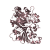
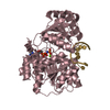
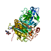
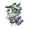
- PDB-1jqg: Crystal Structure of the Carboxypeptidase A from Helicoverpa Armigera -
+ Open data

- Basic information
| Entry | Database: PDB / ID: 1jqg | ||||||
|---|---|---|---|---|---|---|---|
| Title | Crystal Structure of the Carboxypeptidase A from Helicoverpa Armigera | ||||||
 Components Components | carboxypeptidase A | ||||||
 Keywords Keywords | HYDROLASE / PRO-PROTEIN | ||||||
| Function / homology |  Function and homology information Function and homology informationcarboxypeptidase A / metallocarboxypeptidase activity / proteolysis / : / zinc ion binding Similarity search - Function | ||||||
| Biological species |  Helicoverpa armigera (cotton bollworm) Helicoverpa armigera (cotton bollworm) | ||||||
| Method |  X-RAY DIFFRACTION / MOLECULAR REPLACEMENT / Resolution: 2.5 Å X-RAY DIFFRACTION / MOLECULAR REPLACEMENT / Resolution: 2.5 Å | ||||||
 Authors Authors | Estebanez-Perpina, E. / Bayes, A. / Vendrell, J. / Jongsma, M.A. / Bown, D.P. / Gatehouse, J.A. / Huber, R. / Bode, W. / Aviles, F.X. / Reverter, D. | ||||||
 Citation Citation | Journal: J.Mol.Biol. / Year: 2001 Title: Crystal structure of a novel mid-gut procarboxypeptidase from the cotton pest Helicoverpa armigera. Authors: Estebanez-Perpina, E. / Bayes, A. / Vendrell, J. / Jongsma, M.A. / Bown, D.P. / Gatehouse, J.A. / Huber, R. / Bode, W. / Aviles, F.X. / Reverter, D. #1: Journal: Insect Biochem.Mol.Biol. / Year: 1998Title: Midgut carboxypeptidase from Helicoverpa armigera (Lepidoptera: Noctuidae) larvae: enzyme characterisation, cDNA cloning and expression. Authors: Bown, D.P. / Wilkinson, H.S. / Gatehouse, J.A. | ||||||
| History |
|
- Structure visualization
| Structure viewer | Molecule: MolmilJmol/JSmol |
|---|
- Downloads & links
-Download
| PDBx/mmCIF format | 1jqg.cif.gz | 97.9 KB | Display | PDBx/mmCIF format |
|---|---|---|---|---|
| PDB format | pdb1jqg.ent.gz | 72.8 KB | Display | PDB format |
| PDBx/mmJSON format | 1jqg.json.gz | Tree view | PDBx/mmJSON format | |
| Others |  Other downloads Other downloads |
-Validation report
| Arichive directory | https://data.pdbj.org/pub/pdb/validation_reports/jq/1jqgftp://data.pdbj.org/pub/pdb/validation_reports/jq/1jqg | HTTPS FTP |
|---|
-Related structure data
| Related structure data |  1ayeS S: Starting model for refinement |
|---|---|
| Similar structure data |


-Links
 PDBj
PDBj
- Assembly
| Deposited unit | 
| ||||||||
|---|---|---|---|---|---|---|---|---|---|
| 1 |
| ||||||||
| Unit cell |
|
-Components
| #1: Protein | Mass: 48539.023 Da / Num. of mol.: 1 Source method: isolated from a genetically manipulated source Source: (gene. exp.) Helicoverpa armigera (cotton bollworm) / Tissue: MIDGUT / Plasmid: pPIC9 / Production host:  Pichia pastoris (fungus) / Strain (production host): GS115 / References: UniProt: O97389, carboxypeptidase A Pichia pastoris (fungus) / Strain (production host): GS115 / References: UniProt: O97389, carboxypeptidase A |
|---|---|
| #2: Chemical | ChemComp-ZN /   Mass: 65.409 Da / Num. of mol.: 1 / Source method: obtained synthetically / Formula: Zn Mass: 65.409 Da / Num. of mol.: 1 / Source method: obtained synthetically / Formula: Zn |
| #3: Water | ChemComp-HOH /  Mass: 18.015 Da / Num. of mol.: 157 / Source method: isolated from a natural source / Formula: H2O Mass: 18.015 Da / Num. of mol.: 157 / Source method: isolated from a natural source / Formula: H2O |
| Has protein modification | Y |
-Experimental details
-Experiment
| Experiment | Method: X-RAY DIFFRACTION / Number of used crystals: 1 |
|---|
- Sample preparation
| Crystal | Density Matthews: 2.12 Å3/Da / Density % sol: 42.05 % | |||||||||||||||||||||||||||||||||||||||||||||||||
|---|---|---|---|---|---|---|---|---|---|---|---|---|---|---|---|---|---|---|---|---|---|---|---|---|---|---|---|---|---|---|---|---|---|---|---|---|---|---|---|---|---|---|---|---|---|---|---|---|---|---|
| Crystal grow | Temperature: 291 K / Method: vapor diffusion, sitting drop / pH: 5.5 Details: PEG 8000, Na-Acetate, Na-Cacodylate, pH 5.5, VAPOR DIFFUSION, SITTING DROP, temperature 291K | |||||||||||||||||||||||||||||||||||||||||||||||||
| Crystal grow | *PLUS pH: 8 / Method: unknown / Details: used macroseeding | |||||||||||||||||||||||||||||||||||||||||||||||||
| Components of the solutions | *PLUS
|
-Data collection
| Diffraction | Mean temperature: 298 K |
|---|---|
| Diffraction source | Source: ROTATING ANODE / Type: RIGAKU RU300 / Wavelength: 1.5418 Å |
| Detector | Type: MARRESEARCH / Detector: IMAGE PLATE / Date: Jan 16, 2000 |
| Radiation | Monochromator: GRAPHITE / Protocol: SINGLE WAVELENGTH / Monochromatic (M) / Laue (L): M / Scattering type: x-ray |
| Radiation wavelength | Wavelength: 1.5418 Å / Relative weight: 1 |
| Reflection | Resolution: 2.5→10 Å / Num. all: 230224 / Num. obs: 21449 / % possible obs: 93.2 % / Observed criterion σ(F): 2 / Observed criterion σ(I): 2 / Rmerge(I) obs: 0.092 / Net I/σ(I): 15.6 |
| Reflection shell | Resolution: 2.5→2.6 Å / Rmerge(I) obs: 0.034 / Mean I/σ(I) obs: 2.5 / % possible all: 93 |
| Reflection | *PLUS Lowest resolution: 12 Å / Num. measured all: 230224 |
| Reflection shell | *PLUS % possible obs: 93 % / Rmerge(I) obs: 0.344 |
- Processing
| Software |
| |||||||||||||||||||||||||
|---|---|---|---|---|---|---|---|---|---|---|---|---|---|---|---|---|---|---|---|---|---|---|---|---|---|---|
| Refinement | Method to determine structure: MOLECULAR REPLACEMENT Starting model: PDB ENTRY 1AYE Resolution: 2.5→10 Å / Isotropic thermal model: Isotropic / Cross valid method: THROUGHOUT / σ(F): 2 / Stereochemistry target values: Engh & Huber
| |||||||||||||||||||||||||
| Refinement step | Cycle: LAST / Resolution: 2.5→10 Å
| |||||||||||||||||||||||||
| Refine LS restraints |
| |||||||||||||||||||||||||
| LS refinement shell | Resolution: 2.5→2.6 Å
| |||||||||||||||||||||||||
| Refinement | *PLUS Lowest resolution: 12 Å / % reflection Rfree: 7 % | |||||||||||||||||||||||||
| Solvent computation | *PLUS | |||||||||||||||||||||||||
| Displacement parameters | *PLUS | |||||||||||||||||||||||||
| Refine LS restraints | *PLUS Type: x_angle_deg / Dev ideal: 1.5 |
