 Movie
Movie Controller

[English] 日本語
 Yorodumi
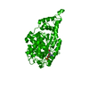
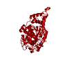
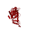
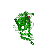
- PDB-1jh8: Structural Investigation of the Biosynthesis of Alternative Lower... -
+ Open data

- Basic information
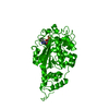
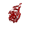
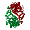
| Entry | Database: PDB / ID: 1jh8 | ||||||
|---|---|---|---|---|---|---|---|
| Title | Structural Investigation of the Biosynthesis of Alternative Lower Ligands for Cobamides by Nicotinate Mononucleotide:5,6-Dimethylbenzimidazole Phosphoribosyltransferase (CobT) from Salmonella enterica | ||||||
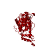
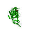
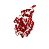
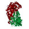
 Components Components | Nicotinate Mononucleotide:5,6-Dimethylbenzimidazole Phosphoribosyltransferase | ||||||
 Keywords Keywords | TRANSFERASE / CobT / Cobalamin biosynthesis / NN:DBI PRT / N1-Alpha-Phosphoribosyltransferase | ||||||
| Function / homology |  Function and homology information Function and homology informationnicotinate-nucleotide-dimethylbenzimidazole phosphoribosyltransferase / nicotinate-nucleotide-dimethylbenzimidazole phosphoribosyltransferase activity / cobalamin biosynthetic process / nucleotide binding Similarity search - Function | ||||||
| Biological species |  Salmonella enterica (bacteria) Salmonella enterica (bacteria) | ||||||
| Method |  X-RAY DIFFRACTION / FOURIER SYNTHESIS / Resolution: 1.8 Å X-RAY DIFFRACTION / FOURIER SYNTHESIS / Resolution: 1.8 Å | ||||||
 Authors Authors | Cheong, C.-G. / Escalante-Semerena, J. / Rayment, I. | ||||||
 Citation Citation | Journal: J.Biol.Chem. / Year: 2001 Title: Structural investigation of the biosynthesis of alternative lower ligands for cobamides by nicotinate mononucleotide: 5,6-dimethylbenzimidazole phosphoribosyltransferase from Salmonella enterica. Authors: Cheong, C.G. / Escalante-Semerena, J.C. / Rayment, I. | ||||||
| History |
|
- Structure visualization
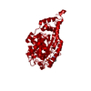
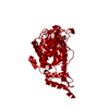
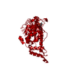
| Structure viewer | Molecule: MolmilJmol/JSmol |
|---|
- Downloads & links
-Download
| PDBx/mmCIF format | 1jh8.cif.gz | 76.5 KB | Display | PDBx/mmCIF format |
|---|---|---|---|---|
| PDB format | pdb1jh8.ent.gz | 56.3 KB | Display | PDB format |
| PDBx/mmJSON format | 1jh8.json.gz | Tree view | PDBx/mmJSON format | |
| Others |  Other downloads Other downloads |
-Validation report
| Arichive directory | https://data.pdbj.org/pub/pdb/validation_reports/jh/1jh8ftp://data.pdbj.org/pub/pdb/validation_reports/jh/1jh8 | HTTPS FTP |
|---|
-Related structure data
| Related structure data |  1jhaC  1jhmC 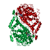 1jhoC  1jhpC  1jhqC  1jhrC  1jhuC  1jhvC  1jhxC  1jhyC  1d0sS S: Starting model for refinement C: citing same article ( |
|---|---|
| Similar structure data |


-Links
 PDBj
PDBj


- Assembly
| Deposited unit | 
| ||||||||
|---|---|---|---|---|---|---|---|---|---|
| 1 | 
| ||||||||
| Unit cell |
|
-Components
| #1: Protein | Mass: 36675.594 Da / Num. of mol.: 1 Source method: isolated from a genetically manipulated source Source: (gene. exp.) Salmonella enterica (bacteria) / Production host: Salmonella enterica (bacteria)References: UniProt: Q05603, nicotinate-nucleotide-dimethylbenzimidazole phosphoribosyltransferase | ||||||
|---|---|---|---|---|---|---|---|
| #2: Chemical |   Mass: 94.971 Da / Num. of mol.: 2 / Source method: obtained synthetically / Formula: PO4 Mass: 94.971 Da / Num. of mol.: 2 / Source method: obtained synthetically / Formula: PO4#3: Chemical | ChemComp-ADE / |   Mass: 135.127 Da / Num. of mol.: 1 / Source method: obtained synthetically / Formula: C5H5N5 Mass: 135.127 Da / Num. of mol.: 1 / Source method: obtained synthetically / Formula: C5H5N5#4: Water | ChemComp-HOH / |  Mass: 18.015 Da / Num. of mol.: 116 / Source method: isolated from a natural source / Formula: H2O Mass: 18.015 Da / Num. of mol.: 116 / Source method: isolated from a natural source / Formula: H2OHas protein modification | Y | |
-Experimental details
-Experiment
| Experiment | Method: X-RAY DIFFRACTION / Number of used crystals: 1 |
|---|
- Sample preparation
| Crystal | Density Matthews: 2.09 Å3/Da / Density % sol: 41.14 % | ||||||||||||||||||||||||||||||
|---|---|---|---|---|---|---|---|---|---|---|---|---|---|---|---|---|---|---|---|---|---|---|---|---|---|---|---|---|---|---|---|
| Crystal grow | Temperature: 298 K / Method: vapor diffusion, hanging drop / pH: 6 Details: 1.4 M Ammonium phosphate, pH 6.0, VAPOR DIFFUSION, HANGING DROP, temperature 298K | ||||||||||||||||||||||||||||||
| Crystal grow | *PLUS | ||||||||||||||||||||||||||||||
| Components of the solutions | *PLUS
|
-Data collection
| Diffraction | Mean temperature: 278 K |
|---|---|
| Diffraction source | Source: ROTATING ANODE / Type: RIGAKU RU200 / Wavelength: 1.54184 |
| Detector | Type: SIEMENS HI-STAR / Detector: AREA DETECTOR / Date: Oct 6, 1999 |
| Radiation | Protocol: SINGLE WAVELENGTH / Monochromatic (M) / Laue (L): M / Scattering type: x-ray |
| Radiation wavelength | Wavelength: 1.54184 Å / Relative weight: 1 |
| Reflection | Resolution: 1.8→30 Å / Num. obs: 25292 / % possible obs: 86.6 % / Observed criterion σ(F): 0 / Redundancy: 2.9 % / Rmerge(I) obs: 0.042 / Net I/σ(I): 15.5 |
| Reflection shell | Resolution: 1.8→1.85 Å / Redundancy: 1.3 % / Rmerge(I) obs: 0.141 / Mean I/σ(I) obs: 2.7 / % possible all: 64.4 |
| Reflection shell | *PLUS % possible obs: 64.4 % |
- Processing
| Software |
| ||||||||||||||||||||
|---|---|---|---|---|---|---|---|---|---|---|---|---|---|---|---|---|---|---|---|---|---|
| Refinement | Method to determine structure: FOURIER SYNTHESIS Starting model: 1D0S Resolution: 1.8→30 Å / Cross valid method: THROUGHOUT / σ(F): 0 / Stereochemistry target values: Engh & Huber
| ||||||||||||||||||||
| Refinement step | Cycle: LAST / Resolution: 1.8→30 Å
| ||||||||||||||||||||
| Refine LS restraints |
| ||||||||||||||||||||
| Software | *PLUS Name: TNT / Classification: refinement | ||||||||||||||||||||
| Refinement | *PLUS Highest resolution: 1.8 Å / Lowest resolution: 30 Å / σ(F): 0 / % reflection Rfree: 10 % / Rfactor obs: 0.175 | ||||||||||||||||||||
| Solvent computation | *PLUS | ||||||||||||||||||||
| Displacement parameters | *PLUS | ||||||||||||||||||||
| Refine LS restraints | *PLUS
|
